CLINICAL CASE
Initial clinical situation
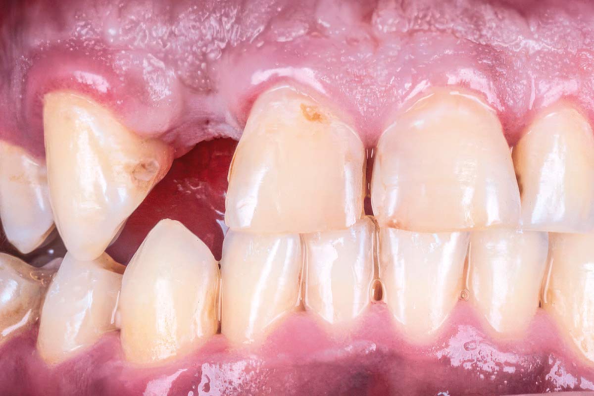
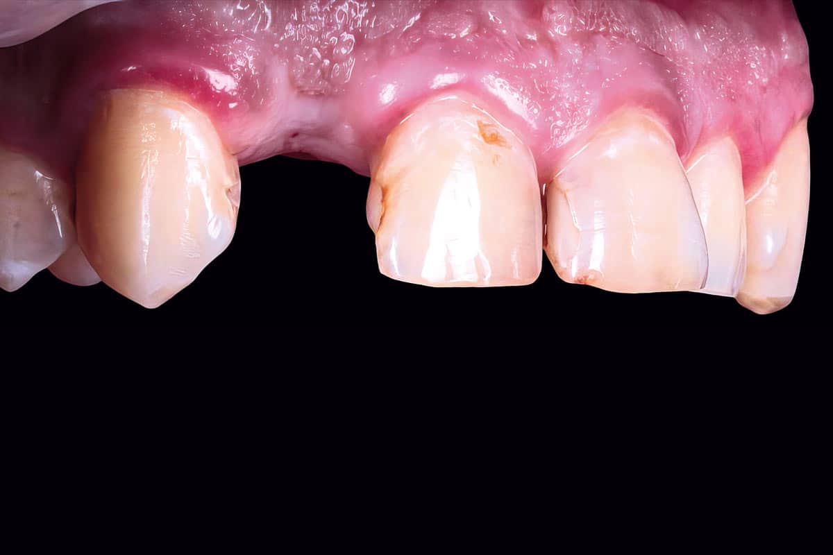
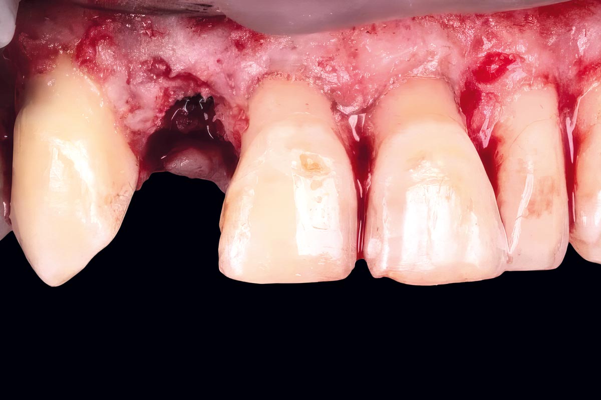
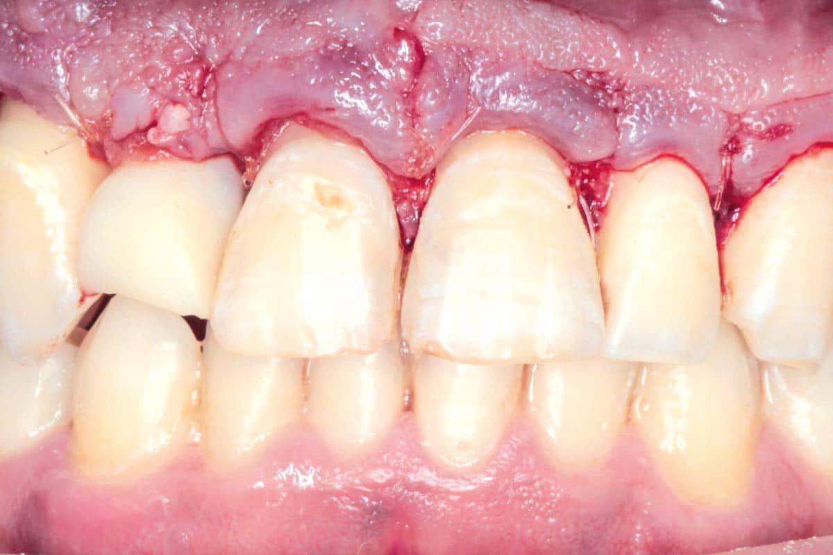
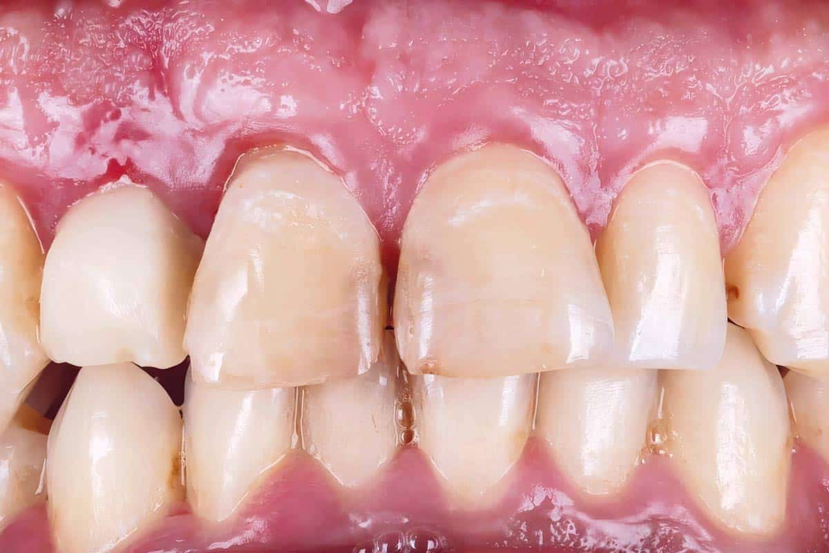
A young adult patient presented with a complex anterior maxillary esthetic situation, including excessive gingival display, short clinical crowns, compromised anterior teeth, and a missing maxillary lateral incisor following previous cystic pathology. Clinical and radiographic evaluation revealed a combined hard and soft tissue defect, with localized collapse of the peri-implant contour and disharmony of the gingival architecture.
The main challenge was not only implant placement, but the reconstruction of a stable and natural soft tissue volume in the esthetic zone. For this reason, mucoderm® was selected as the central biomaterial to enhance peri-implant soft tissue thickness and contour while avoiding palatal donor site morbidity.
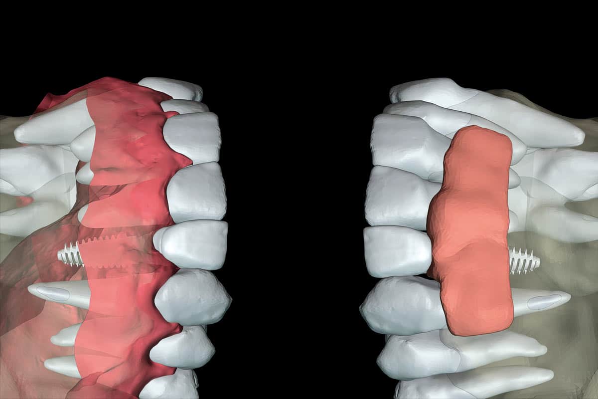
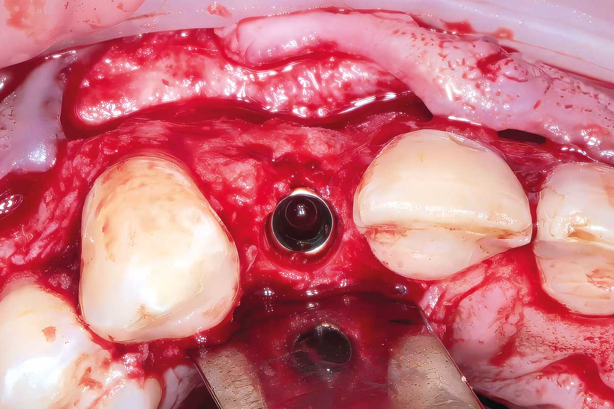
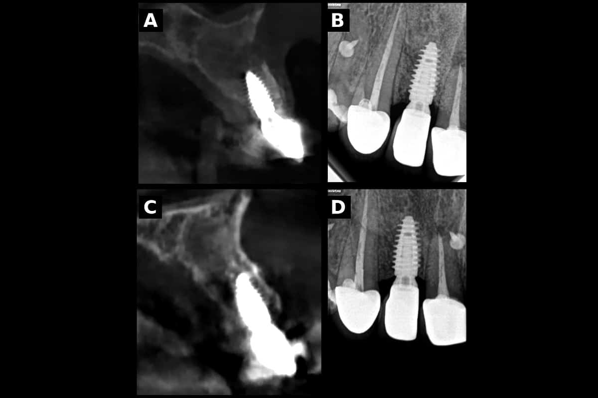
The case was planned digitally, defining the final crown proportions, gingival margins, implant position, and required soft-tissue volume before surgery. A guided crown-lengthening procedure with vertical osseous recontouring was first performed to correct the gingival architecture and establish a stable hard-tissue foundation. The implant was then placed in a prosthetically driven position at the lateral incisor site.
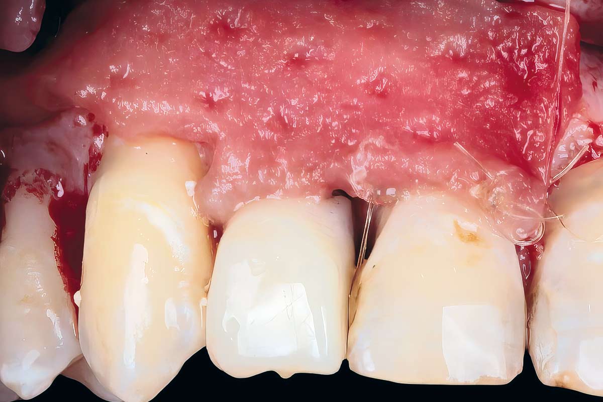
The soft tissue deficiency was reconstructed using mucoderm®. The matrix was shaped extraorally with the help of a 3D-printed guide derived from the digital plan, allowing patient-specific adaptation to the defect morphology. The customized mucoderm® was then positioned and stabilized using a poncho-type approach beneath the immediate provisional restoration, which helped support and condition the healing soft tissue.
After uneventful healing and tissue maturation, the definitive restorations were delivered using layered zirconia.
Clinical outcomes
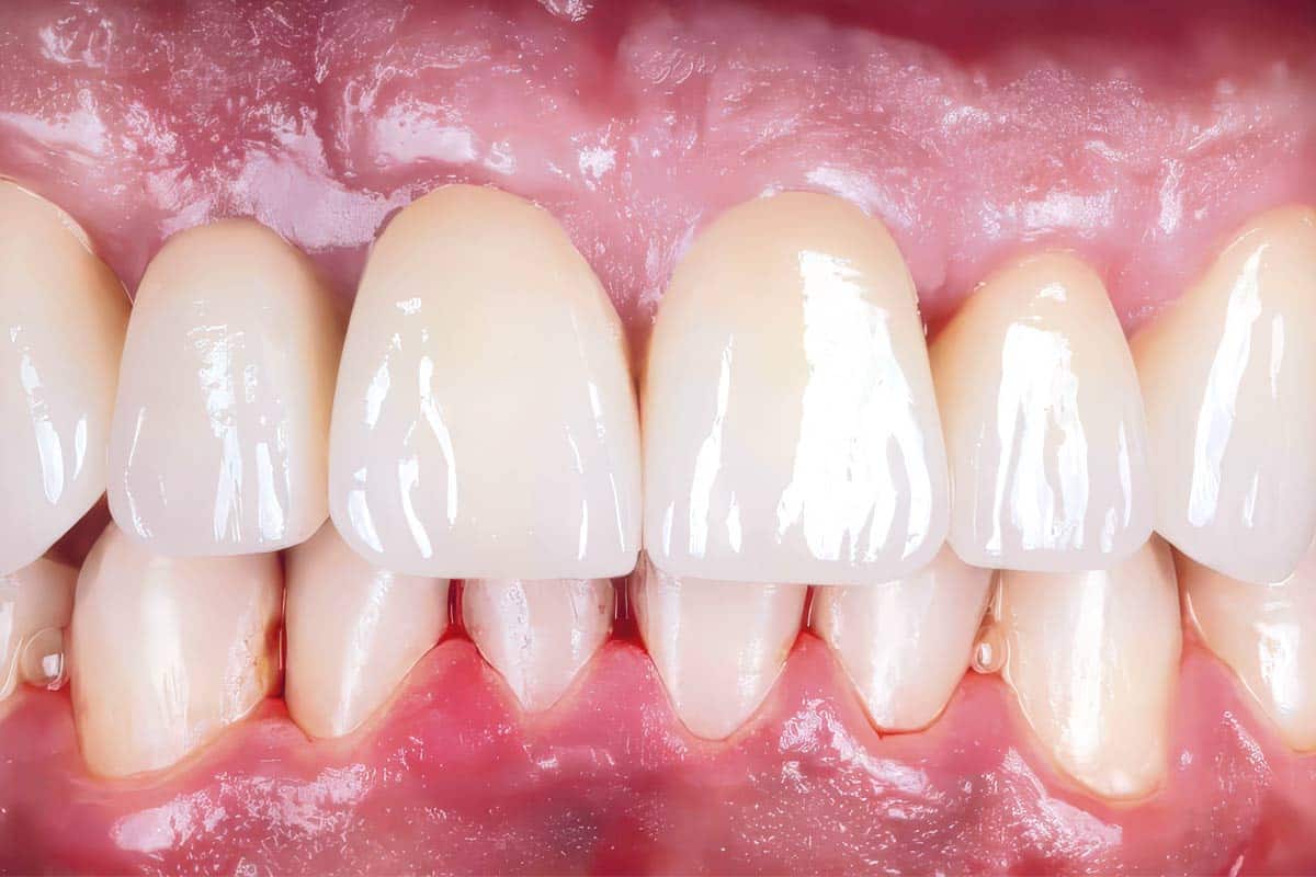
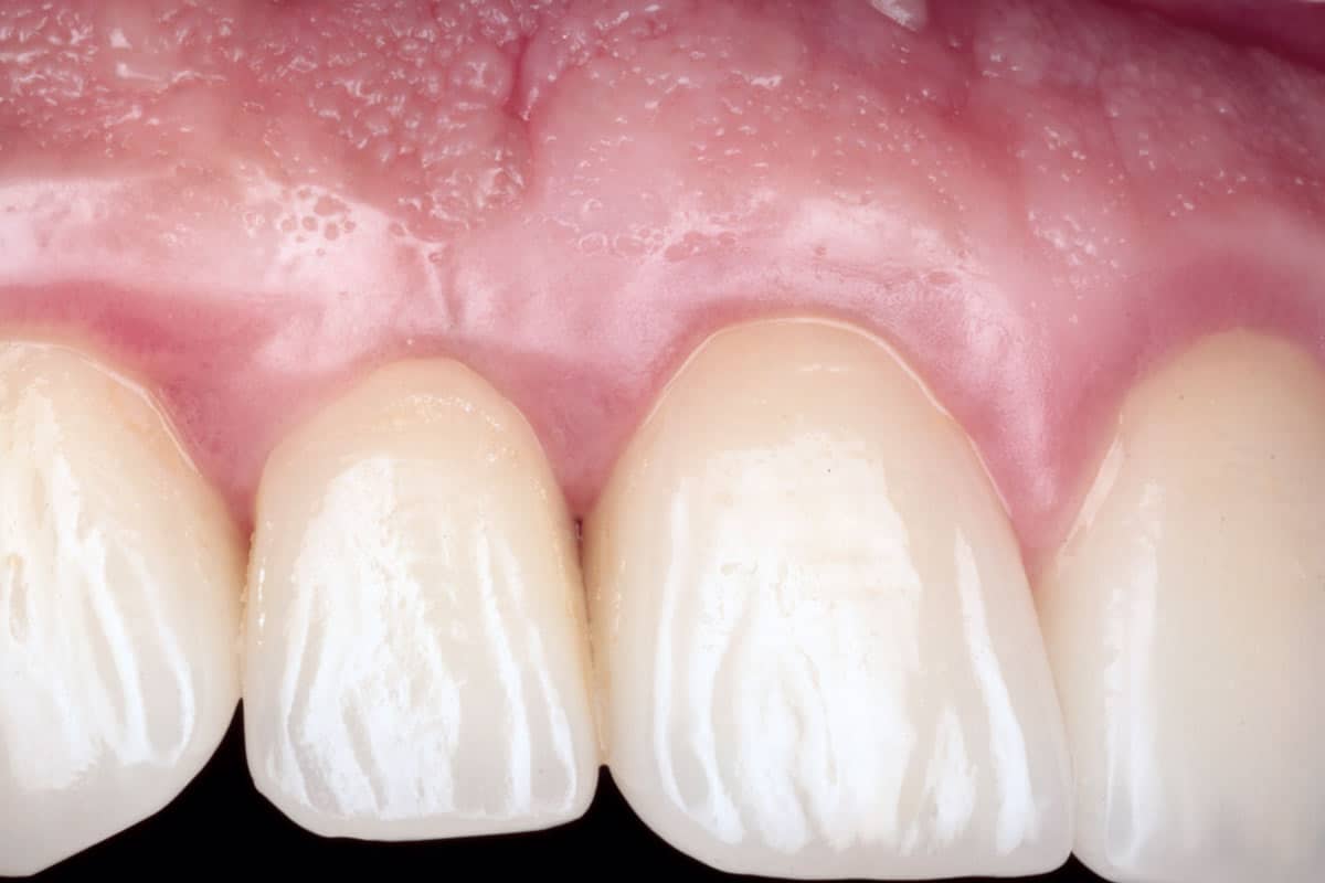
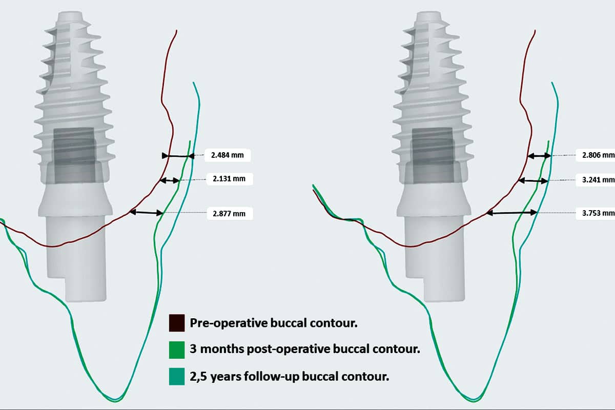
The mucoderm® graft contributed to a clear improvement in peri-implant soft-tissue volume, creating a more convex and harmonious buccal contour at the implant site. The reconstructed tissue showed improved transition with the adjacent teeth and supported a natural emergence profile for the final restoration.
At follow-up, the clinical outcome demonstrated stable soft-tissue maturation, preservation of the reconstructed contour, and harmonious integration of the implant-supported restoration within the anterior maxilla. The digital contour analysis further supported the clinical observation of progressive buccal soft-tissue thickening and stable buccal convexity over time.
Key results included improved peri-implant soft-tissue thickness, enhanced buccal convexity, stable esthetic integration, reduced patient morbidity by avoiding palatal harvesting, and successful use of mucoderm® as a digitally planned patient-specific soft-tissue scaffold.
Conclusion
This case highlights mucoderm® as a key component in the reconstruction of peri-implant soft-tissue volume in a challenging esthetic-zone defect. When combined with digital planning, guided surgery, immediate provisionalization, and patient-specific matrix shaping, mucoderm® allowed a controlled, minimally invasive, and esthetically stable soft-tissue augmentation.