KLINISCHER FALL
Dr. Narissaporn Chaiprakit
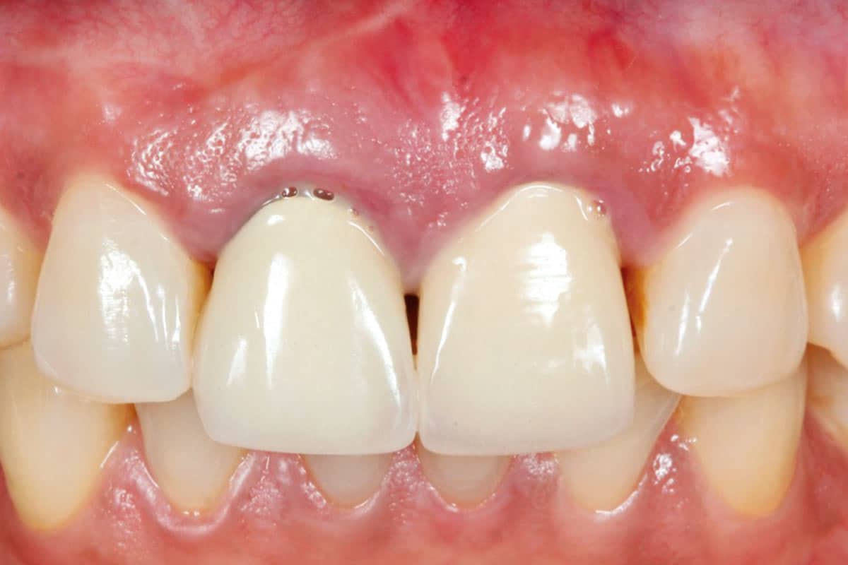
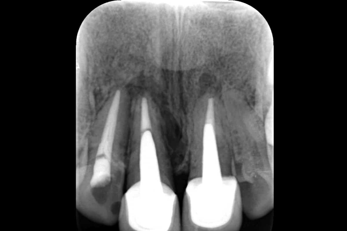
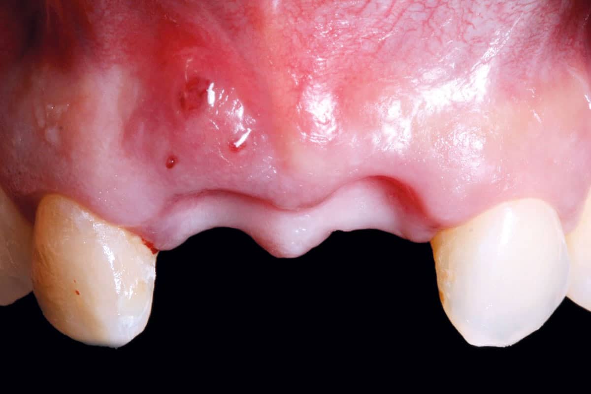
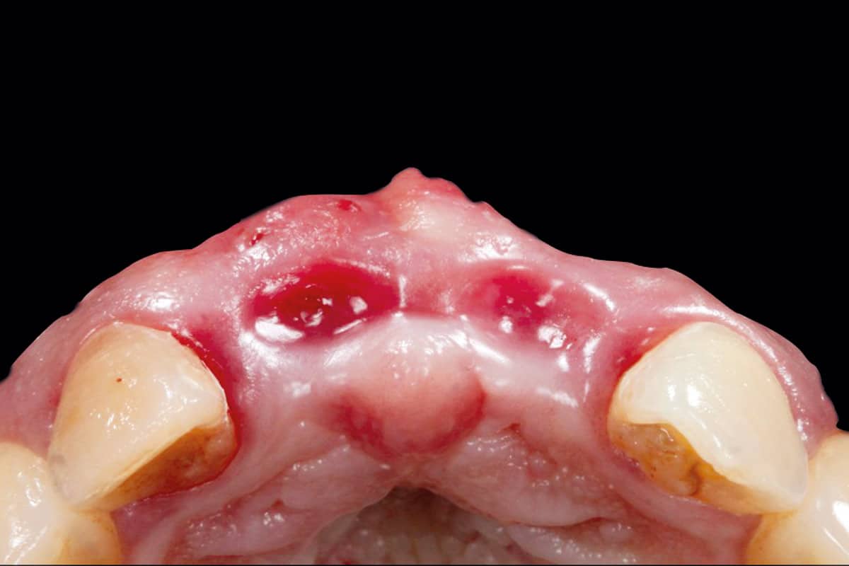
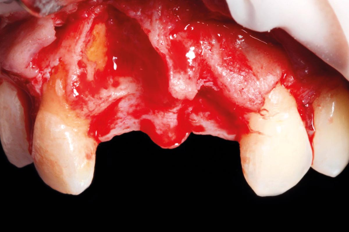
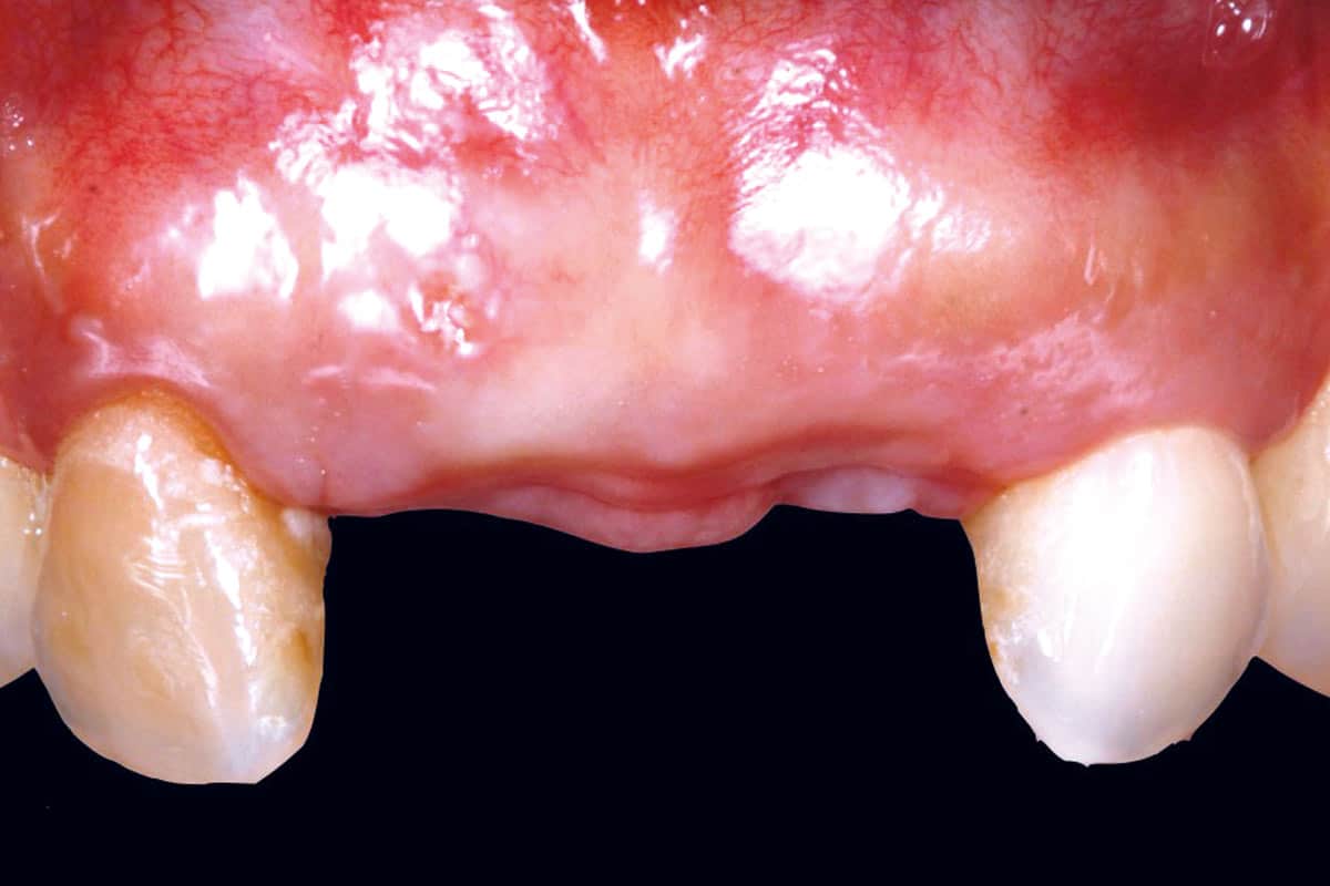
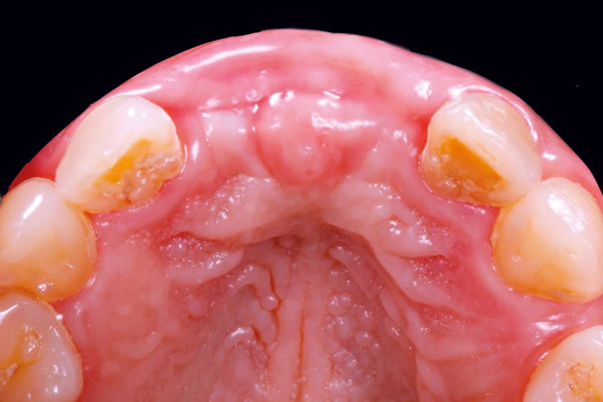
The patient presented with compromised maxillary central incisors 11 and 21 confirmed also radiographically. Following extraction, the sites were allowed to heal for 8 weeks. Clinical examination revealed a pronounced loss of ridge volume, particularly on the facial aspect.
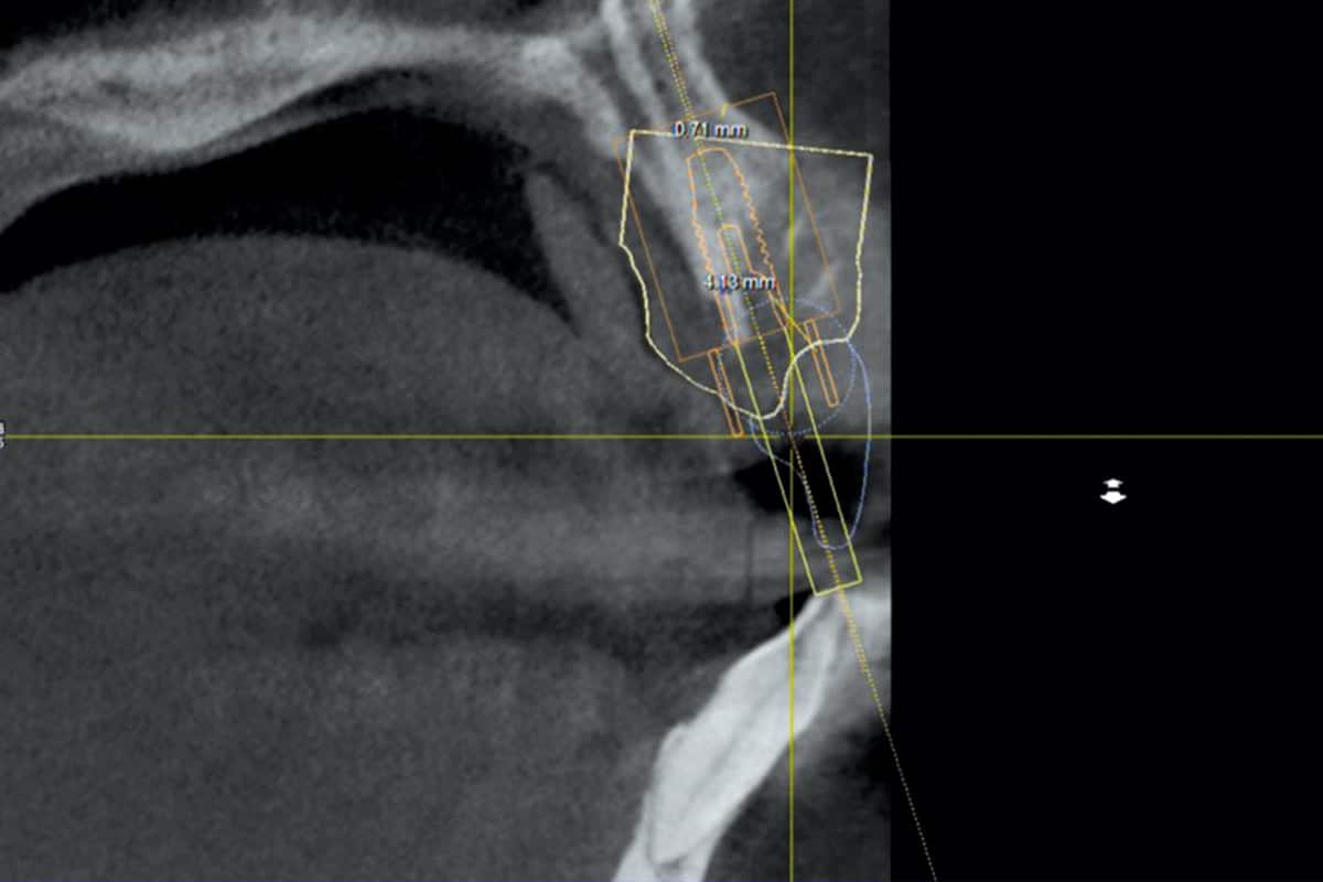
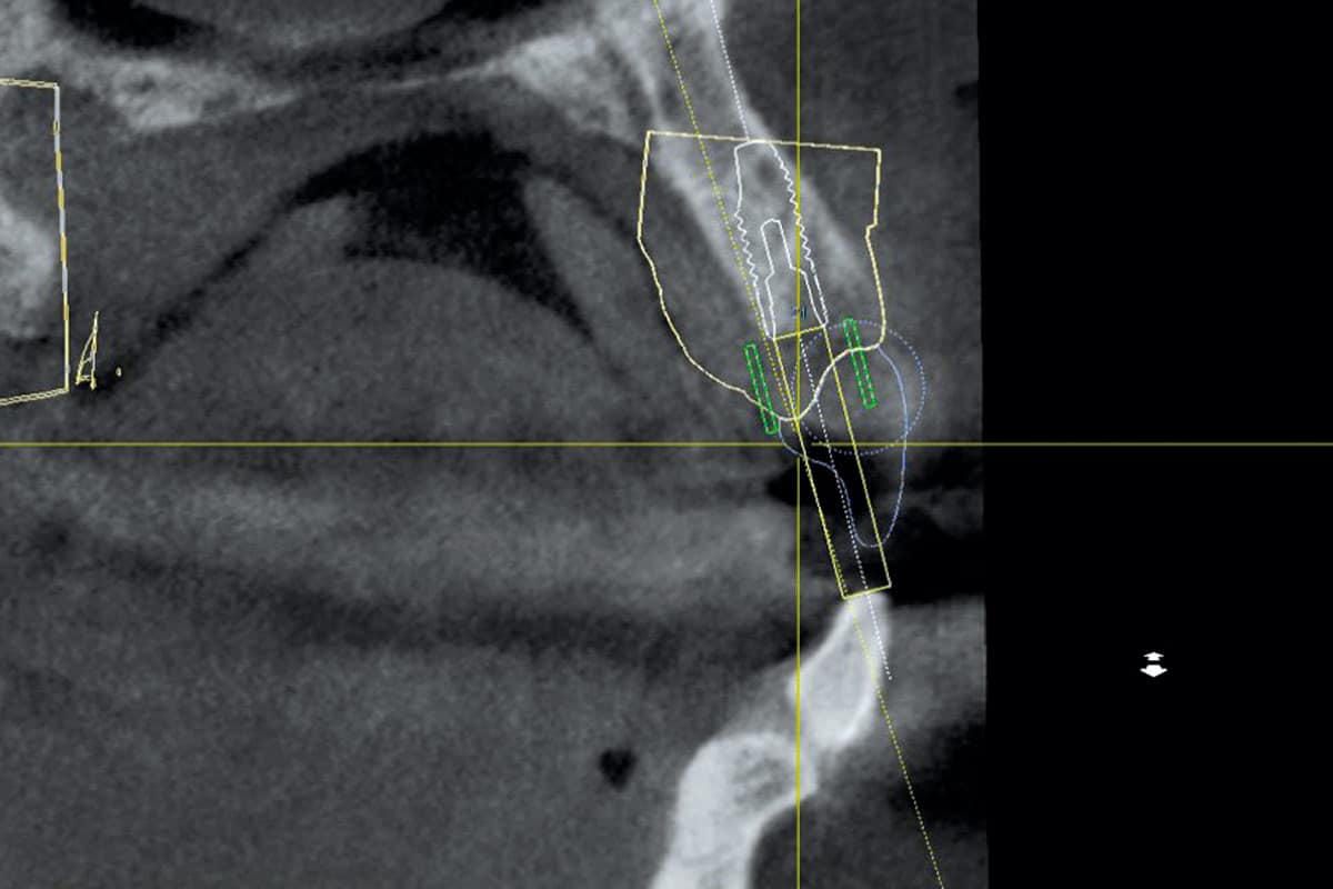
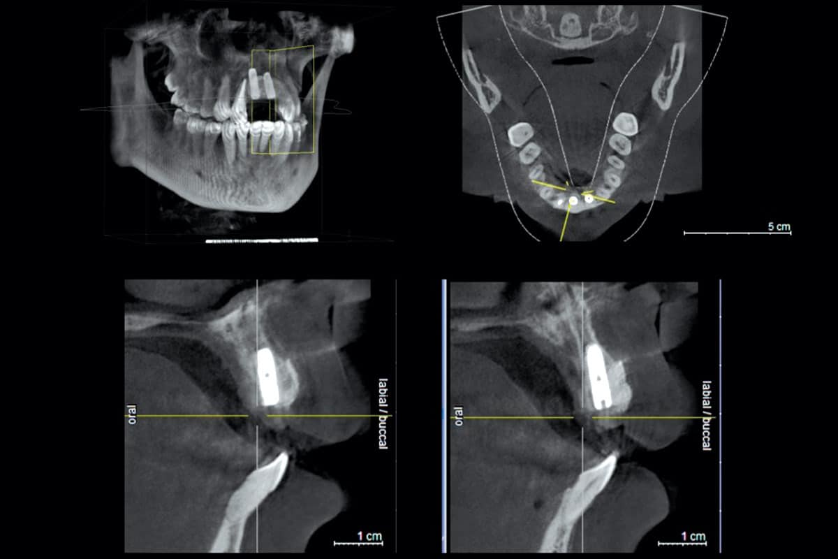
CBCT analysis demonstrated insufficient facial bone volume, confirming the need for simultaneous horizontal augmentation. Following flap elevation, the extent of the osseous defect was clinically confirmed.
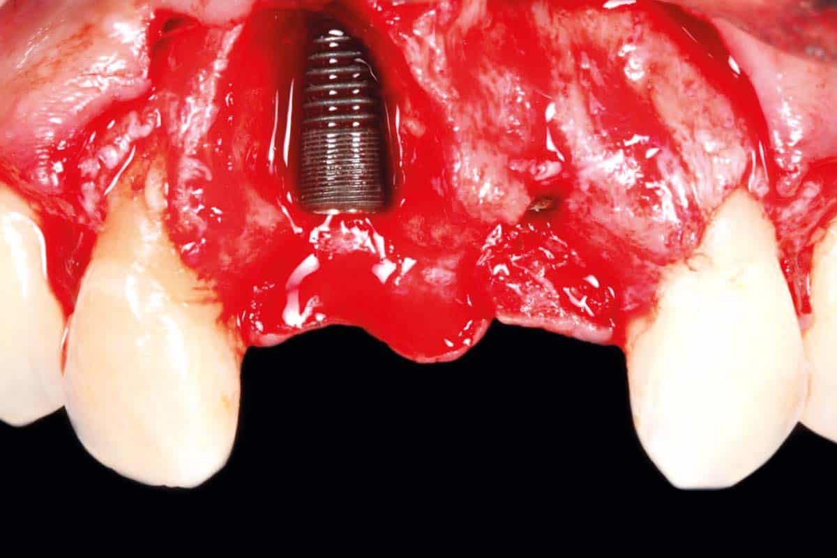
Implant placement and simultaneous GBR:
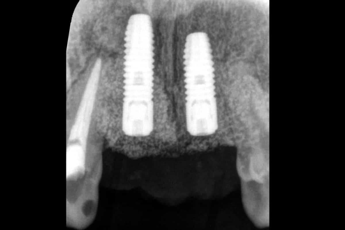
Two implants were placed.
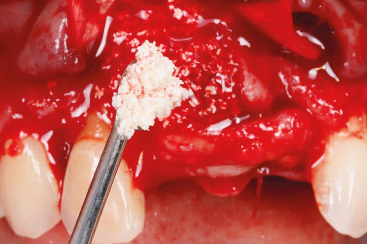
The deficient facial contour was reconstructed using a combination of autogenous bone and cerabone®. The augmented area was subsequently covered with Jason® membrane to provide barrier function and maintain separation between the regenerative compartment and the overlying soft tissues.
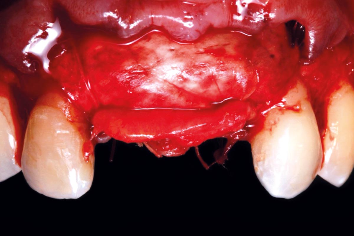
A connective tissue graft (CTG) was additionally placed to increase soft-tissue volume and support the peri-implant tissue architecture in the esthetic zone.
Healing and prosthetic rehabilitation:
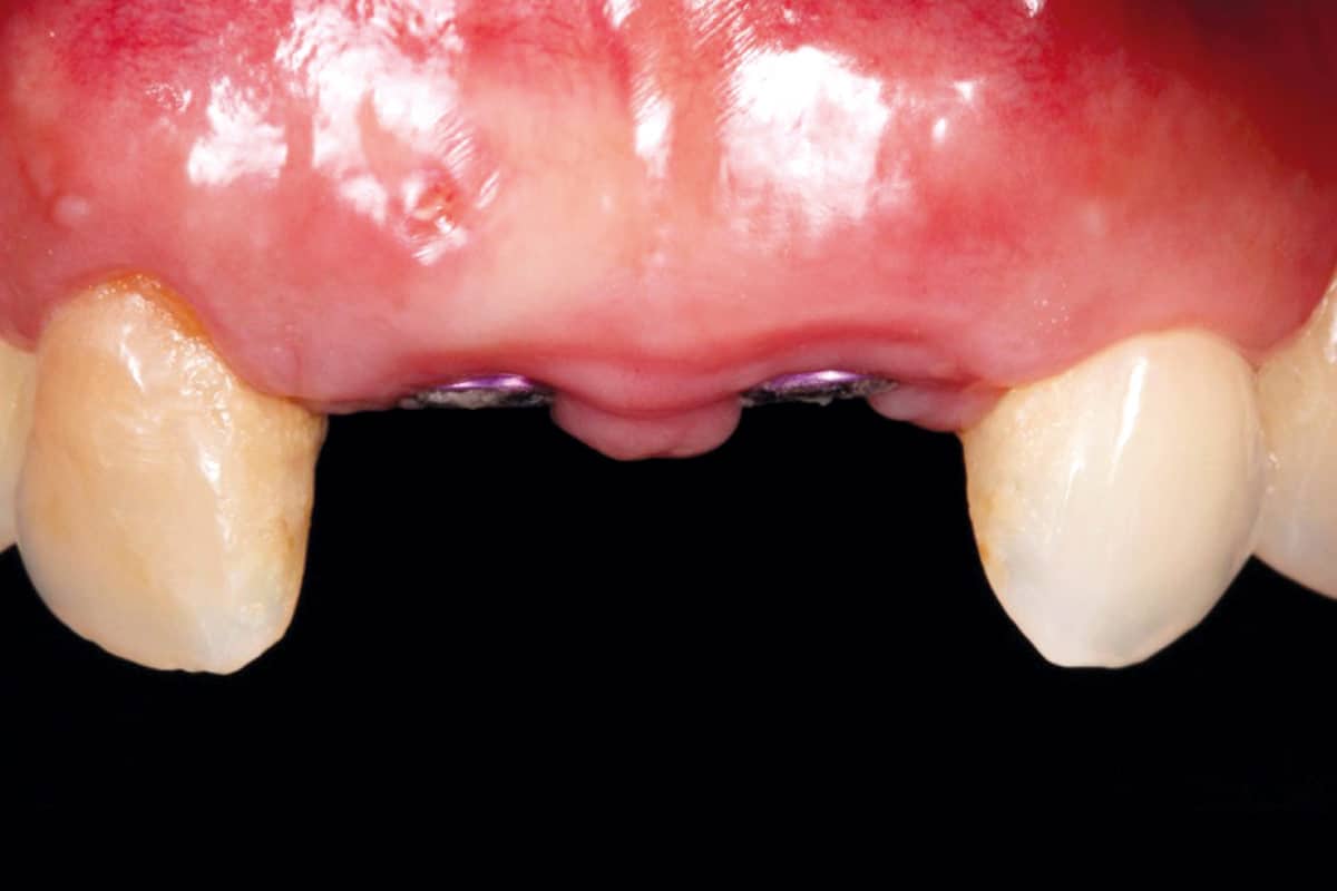
After 4 months of healing, clinical and radiographic evaluation demonstrated successful reconstruction of the anterior ridge with favorable hard- and soft-tissue conditions.
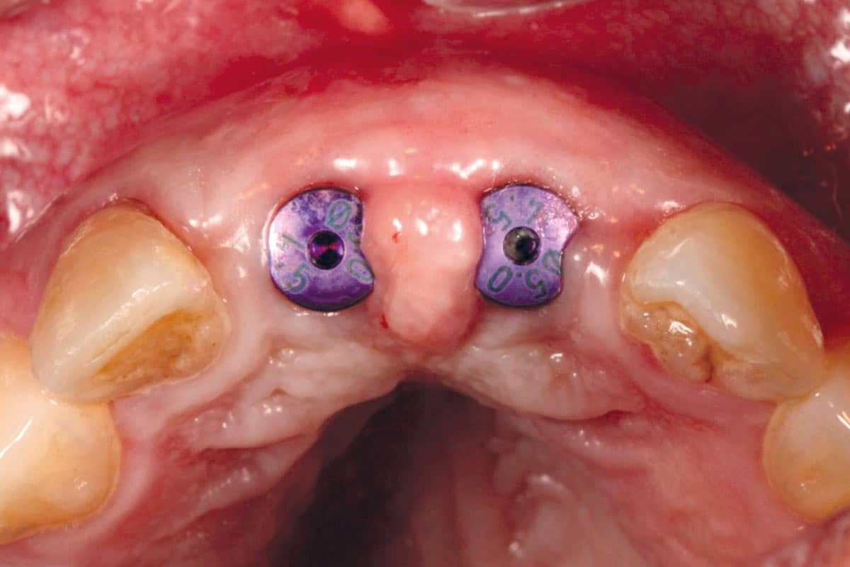
At approximately 5 months, the implants were uncovered and the prosthetic phase was initiated. Temporary restorations were used to condition the peri-implant soft tissues and establish an appropriate emergence profile prior to definitive rehabilitation.
Long-term outcome:
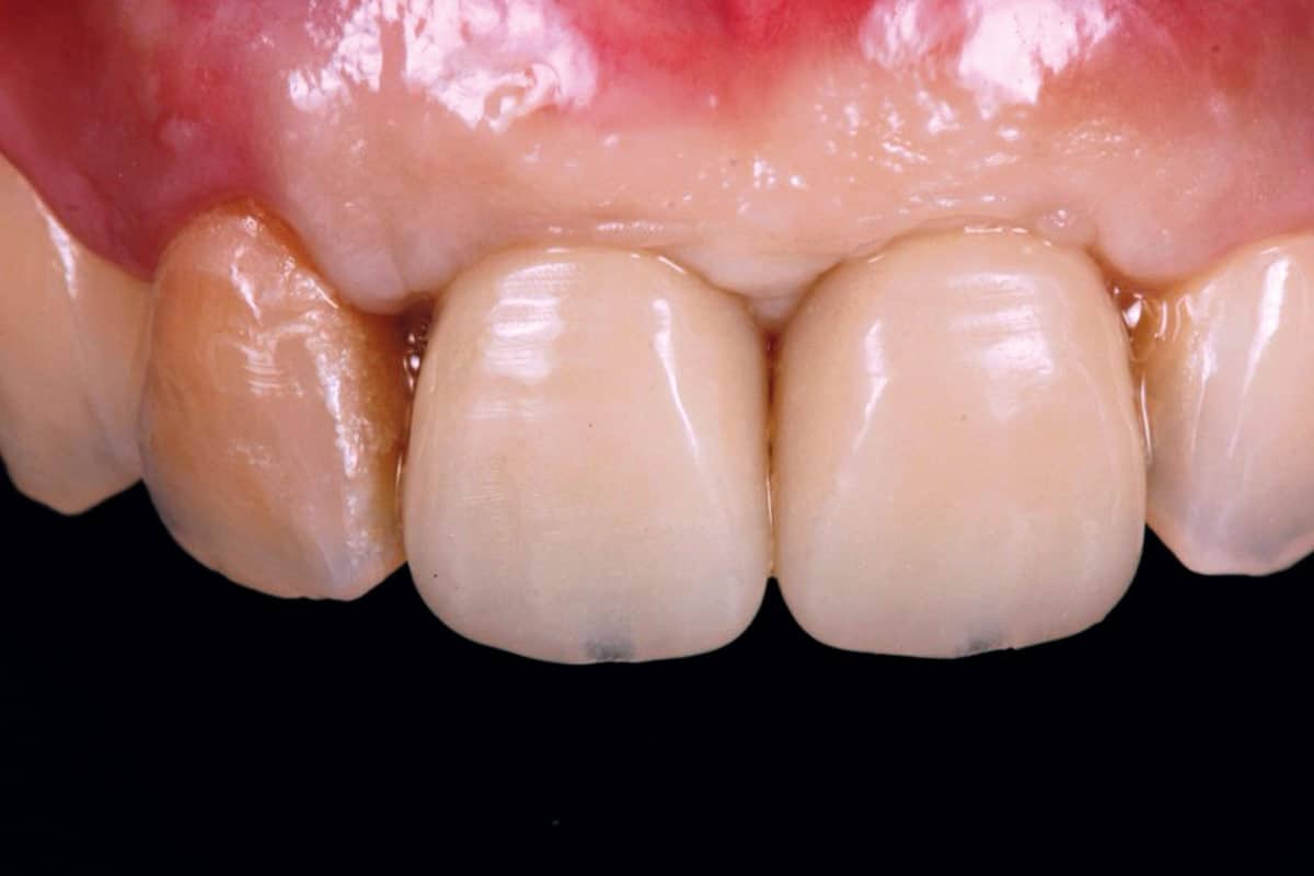
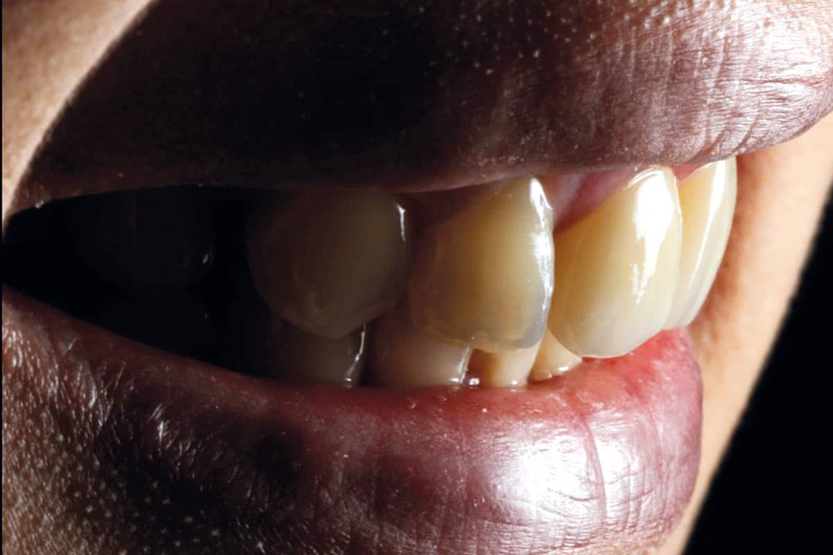
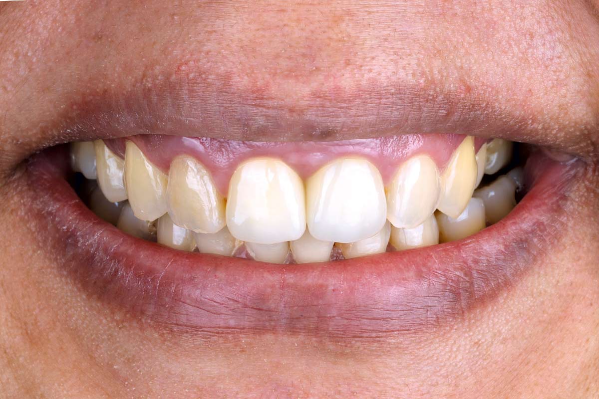
At the 1-year follow-up, the implant-supported restorations showed favorable integration and stable peri-implant tissue contours.
Most importantly, the 8-year follow-up demonstrates maintenance of the clinical and esthetic outcome, highlighting the long-term stability of the reconstructed anterior ridge.