Studie
Alveolar ridge and keratinized gingiva preservation using mucoderm® and cerabone® in flapless extractions
Original Title: Alveolar ridge and keratinized gingiva preservation using collagen matrix and inorganic bone substitute in flapless extractions: A case series of exposed biomaterials
Mariana Moraes de Souza, Cristhiam de Jesus Hernandez Martinez, Valessa Florindo Carvalho, Flávia Aparecida Chaves Furlaneto Messora, Daniela Bazan Palioto Bull, Michel Reis Messora, Sérgio Luís Scombatti de Souza, Arthur Belém Novaes Jr, Mario Taba Jr
Journal of the International Academy of Periodontology (2022) 24/4: 367-77
Read full study here
This is the first controlled study to evaluate the dimensional changes after alveolar ridge preservation using cerabone® and intentionally left exposed mucoderm® with flapless and flap approach.
Aim
The aim of the clinical randomized case series was to evaluate the dimensional changes following an alveolar ridge preservation procedure comparing flapless and flap techniques in combination with bovine bone mineral (cerabone®) and a collagen matrix (mucoderm®) intentionally left exposed.
Methods
Systematically healthy patients with a need for extraction of premolars and present tooth adjacent to the extraction site were selected.
Patients were randomly treated with either flapless technique (Group 1, G1) or with intrasulcular and oblique incisions on the distal surface of the adjacent tooth, performed with a total flap (Group 2, G2).
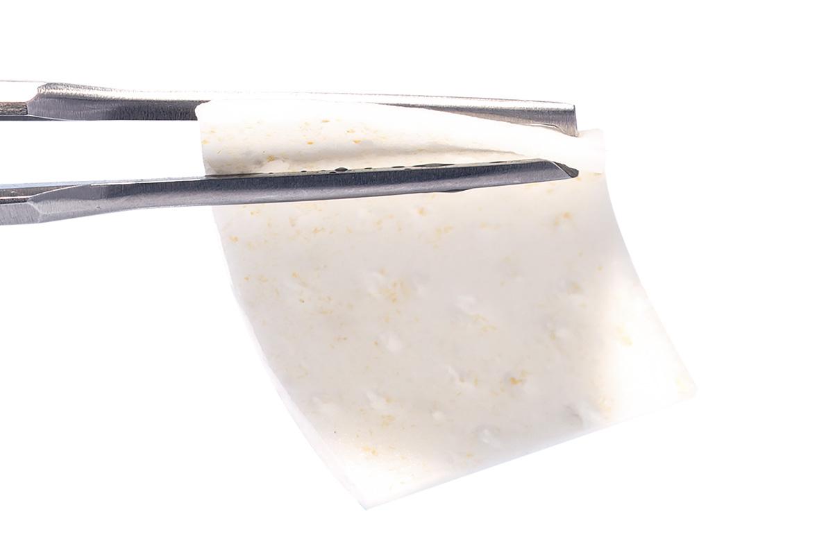
All sockets were filled with cerabone® granules and covered with mucoderm® intentionally left exposed. Simple suturing was performed in order to stabilize mucoderm® in G1, whereby in G2 the flap was repositioned and sutured in the original position of the gingival margin.
Post-operatively clinical and CBCT measurements were carried out at 1 week, 4 months and 2 years. Radiological analysis included assessment of dimensional changes of the ridge, i.e. the buccal and lingual/palatal cortical bone height and the horizontal width of the extraction socket (measured at 1 mm, 3 mm, and 5 mm below the lingual bone crest). Clinical analysis assessed keratinized gingiva height and thickness. Furthermore, postoperative discomfort was evaluated using a Visual Analog Scale (VAS) patient diary.
Results
Results on soft tissue regeneration
For the clinical parameters (height and thickness of keratinized gingiva) there were no statistical differences between the two groups at the different evaluation times. However, the flapless approach seems to have slightly more favorable results in the changes of the height and thickness of the keratinized gingiva after 4 and 24 months compared to the flap approach.
Results on hard tissue regeneration
- Both groups showed reduction in buccal and lingual height, as well as in horizontal width of the extraction socket, but there were no statistically significant differences. However, the flapless approach (G1) showed smaller losses than flap approach (G2) in the radiological parameters after 4 months and 2 years.
- Overall, this study found limited bone resorption in the horizontal width of the socket below the marginal bone crest, when compared to spontaneous healing in other studies and systematic reviews.
Patient-related outcomes
- During the first 7 days post-operatively statistically significant difference in VAS in favor of the flapless group was reported (5.53 ± 9.68) indicating less pain and discomfort for the flapless group as compared to the flap approach (15.31 ± 21.08), however no statistically significant difference for the other periods was found.
- Surgical procedures were performed uneventfully and no healing complications were reported.
Results on biomaterials used
- cerabone® and mucoderm® have been demonstrated to perform efficiently in ridge preservation, both with flapless and flap techniques.
- mucoderm® was intentionally left exposed and served to support soft and hard tissue regeneration as no signs of infection or necrosis of both biomaterials was observed.
Conclusion
- Alveolar ridge preservation limited vertical and horizontal socket changes, regardless of the surgical technique used.
- The flapless approach provided significantly less discomfort in the initial healing phase and seemed advantageous regarding ridge dimensional changes.


