Studie
In vitro comparison of the osteogenic capability of human pulp stem cells on alloplastic, allogeneic, and xenogeneic bone scaffolds
>https://pubmed.ncbi.nlm.nih.gov/36721114/
Heitzer M, Modabber A, Zhang X, et al. BMC Oral Health. 2023;23(1):56. Published 2023 Jan 31. doi:10.1186/s12903-023-02726-4
This in vitro study investigates the osteogenic capability of dental pulp stem cells on alloplastic (maxresorb®), allogeneic (maxgraft®), and xenogeneic (cerabone®) bone grafts. The aim of this study was to determine the favorable amount of scaffolding for stem cell cultivation to be used for augmentative procedures of maxillomandibular bone defects.
Background:
A rigorous search for alternatives to autogenous bone grafts to avoid invasiveness at the donor site in the treatment of maxillomandibular bone defects. Researchers have used alloplastic, allogeneic, and xenogeneic bone graft substitutes in clinical studies with varying degrees of success, although their in vitro effects on stem cells remain unclear. Dental pulp stem cells (DPSCs) can potentially enhance the bone regeneration of bone graft substitutes. The present in vitro study investigates the osteogenic capability of DPSCs on alloplastic (biphasic calcium phosphate [BCP]), allogeneic (freeze-dried bone allografts [FDBAs]), and xenogeneic (deproteinized bovine bone mineral [DBBM]) bone grafts. The optimal amounts of BCP, DBBM, and FDBA were investigated in relation to the proliferation, viability, attachment, and osteogenic differentiation of DPSC in vitro.
Methods:
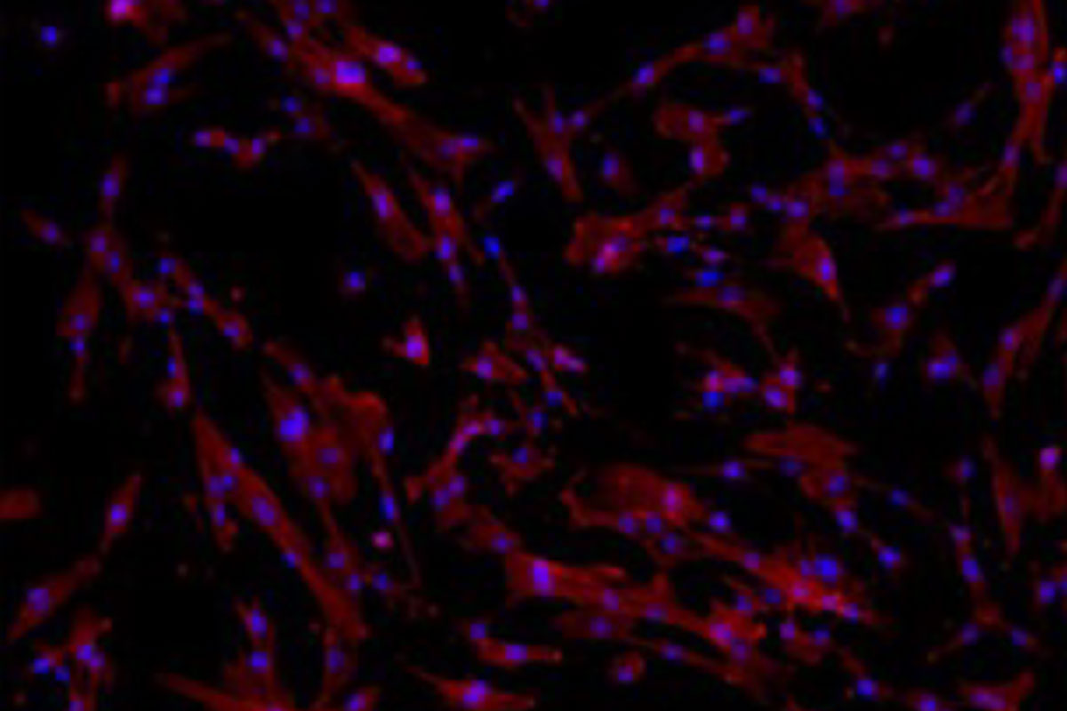
Human DPSCs were seeded on 0.5 mg/ml, 1 mg/ml, and 2 mg/ml of BCP, FDBA, and DBBM to evaluate the optimal cell growth and cytotoxicity. Scaffolds and cell morphologies were analyzed by scanning electron microscopy (SEM). Calcein AM and cytoskeleton staining were performed to determine cell attachment and proliferation. Alkaline phosphatase (ALP) and osteogenesis-related genes expressions was used to investigate initial osteogenic differentiation.
Results:
Cytotoxicity assays showed that most viable DPSCs were present at a scaffold concentration of 0.5 mg/ml. The DPSCs on the cerabone® scaffold demonstrated a significantly higher proliferation rate of 214.25 ± 16.17 (p < 0.001) cells, enhancing ALP activity level and upregulating of osteogenesis-related genes compared with other two scaffolds.
Conclusion:
This study has shown the suitability of maxresorb®, maxgraft®, and cerabone® for colonization with human dental pulp stem cells (DPSCs) for tissue engineering applications, especially for the treatment of maxillomandibular bone defects. The deproteinized bovine bone mineral (DBBM) cerabone® in particular showed the most favorable properties in terms of proliferation, attachment, and osteogenic differentiation.