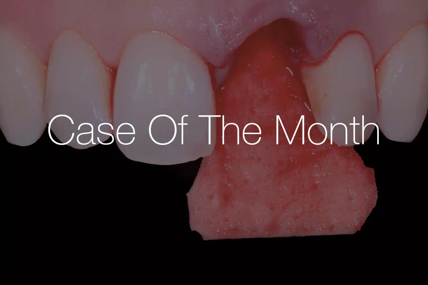

CASE OF THE MONTH | 03/2026
Dr. Alessandro Rossi
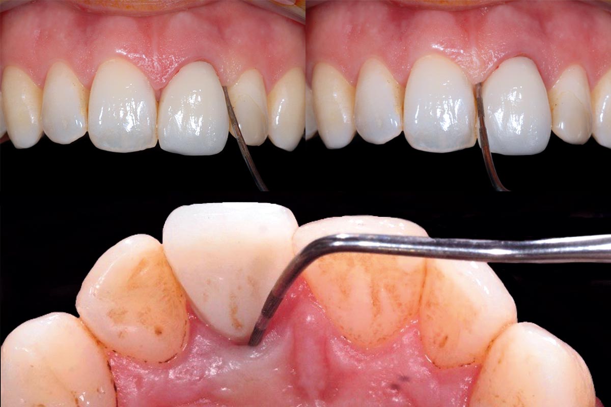
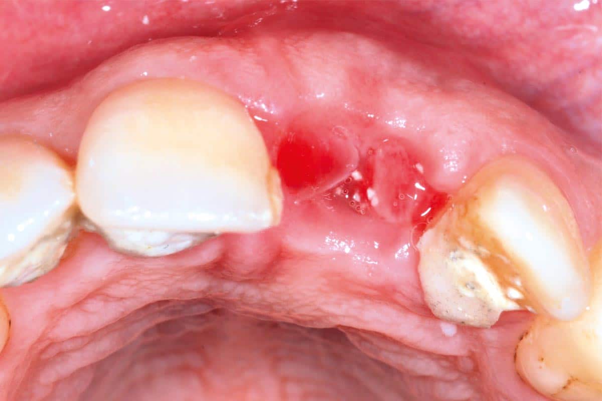
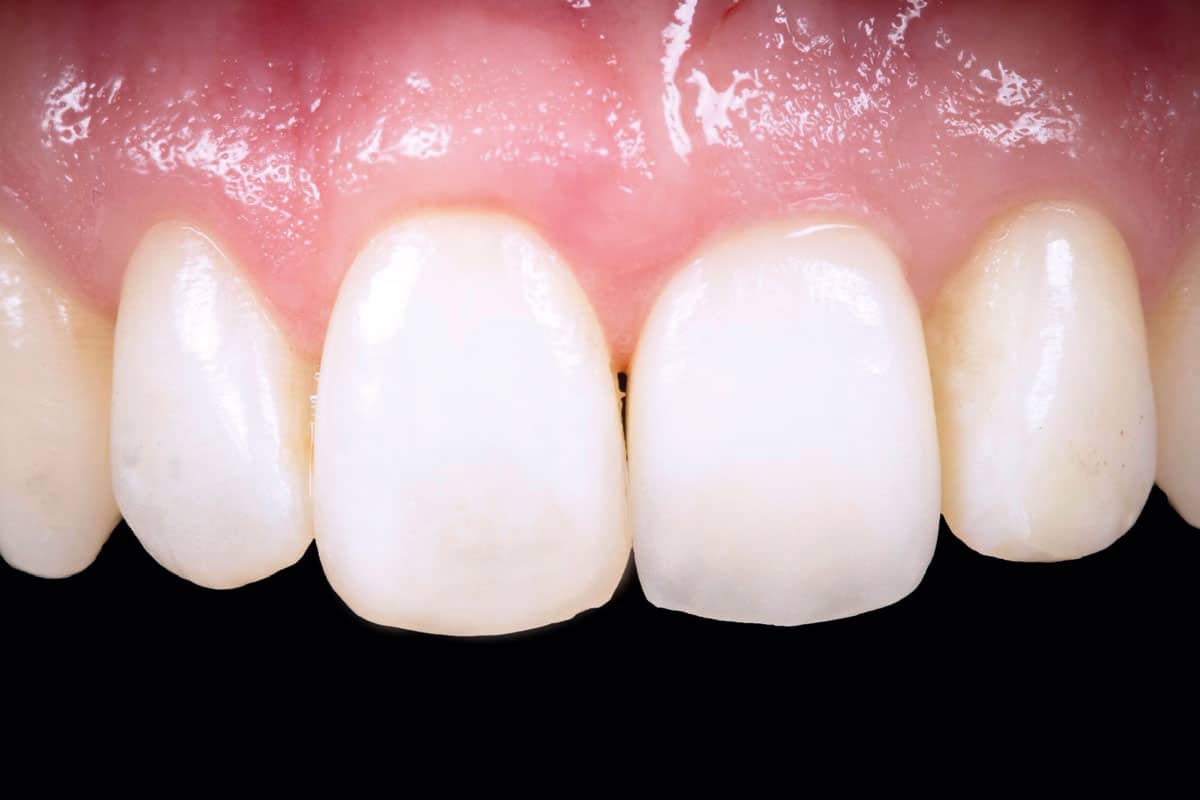
The patient presented with a single-rooted tooth indicated for extraction. The Periodontal charting of the site showed a probing dept of 10 mm only on the palatal aspect, compatible with the diagnosis of vertical fracture. The periapical radiograph demonstrated deep root decay with associated hard tissue destruction.
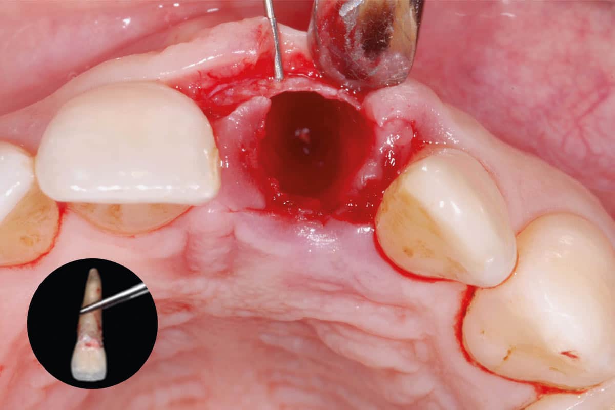
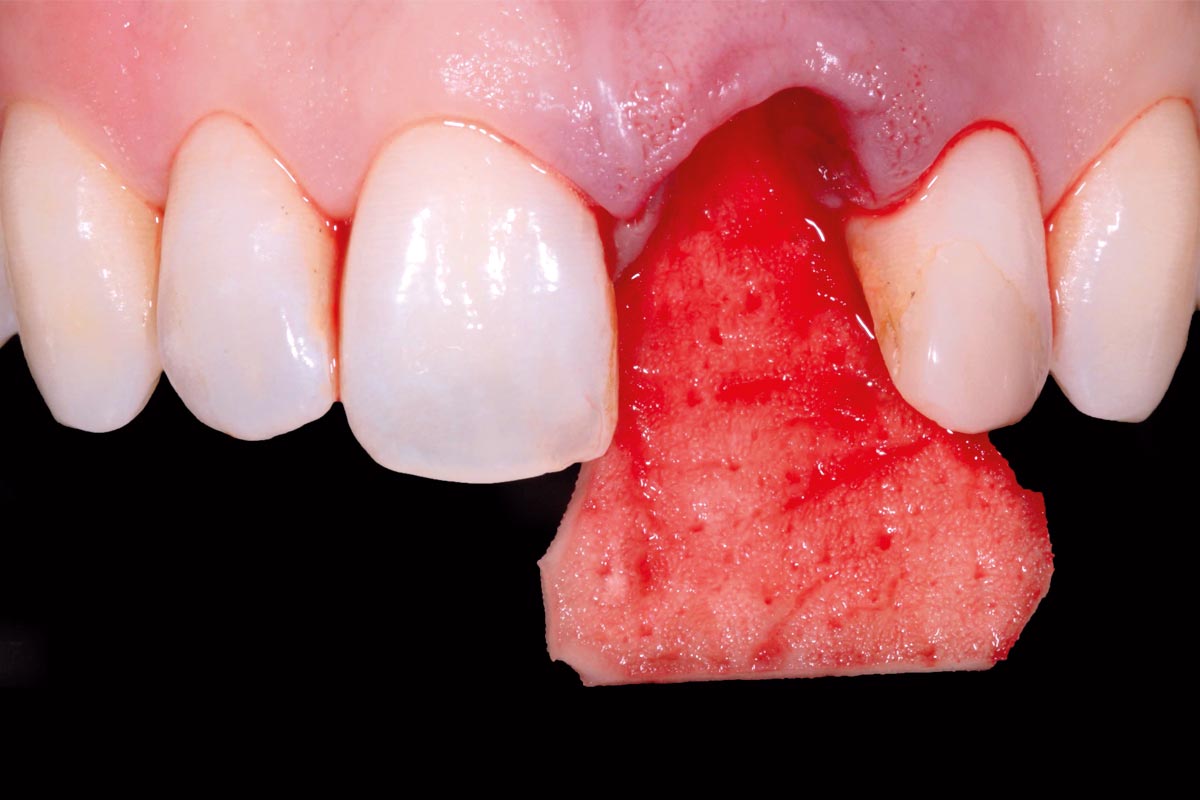
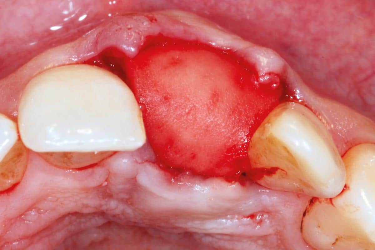
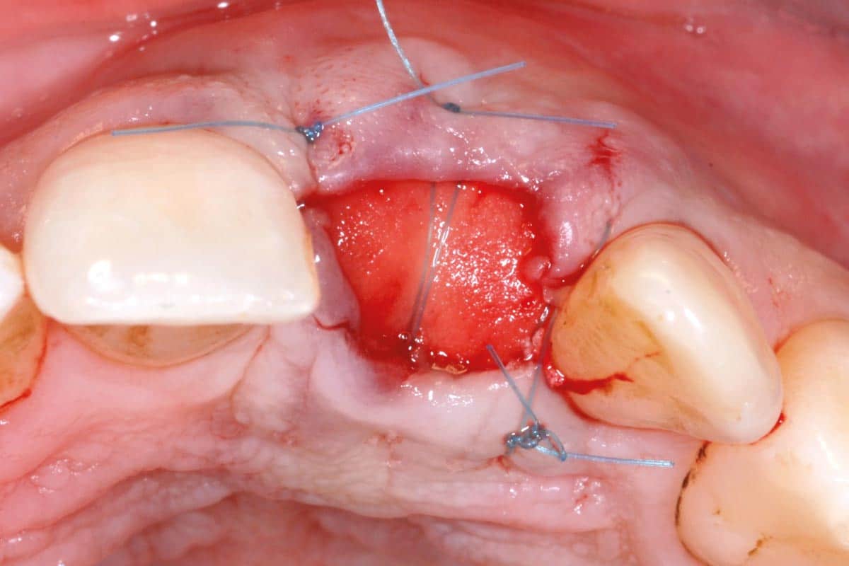
The tooth was removed atraumatically, and on the palatal aspect of the root was evidenced a vertical fracture. The extraction socket was subsequently thoroughly debrided to eliminate granulation tissue and ensure a clean recipient site. Intraoperative assessment revealed a buccal bone thickness of ≤ 1 mm. To preserve alveolar ridge dimensions and prepare the site for a staged implant approach, the socket was grafted with a bone substitute material for ridge preservation. For simultaneous soft tissue augmentation and socket sealing, mucoderm®, was used in a combined onlay–interpositional technique. The buccal and palatal portions of the matrix were inserted beneath the respective full-thickness flaps, while the central portion was positioned over the crestal aspect of the socket and intentionally left exposed to the oral cavity to achieve socket sealing. Wound closure was accomplished with interrupted sutures placed interproximally and an internal horizontal mattress suture connecting the mid-facial and mid-palatal aspects of the flaps to ensure stable fixation and gentle compression of the matrix.
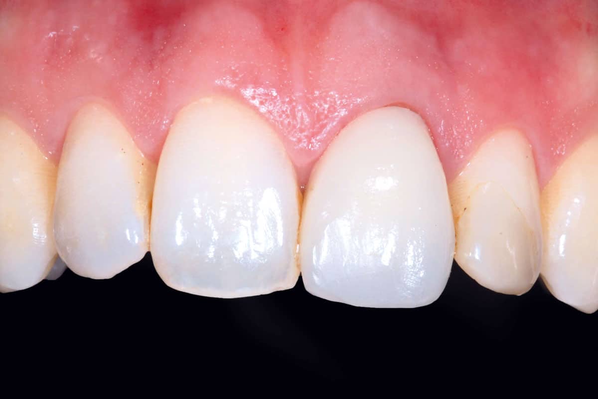
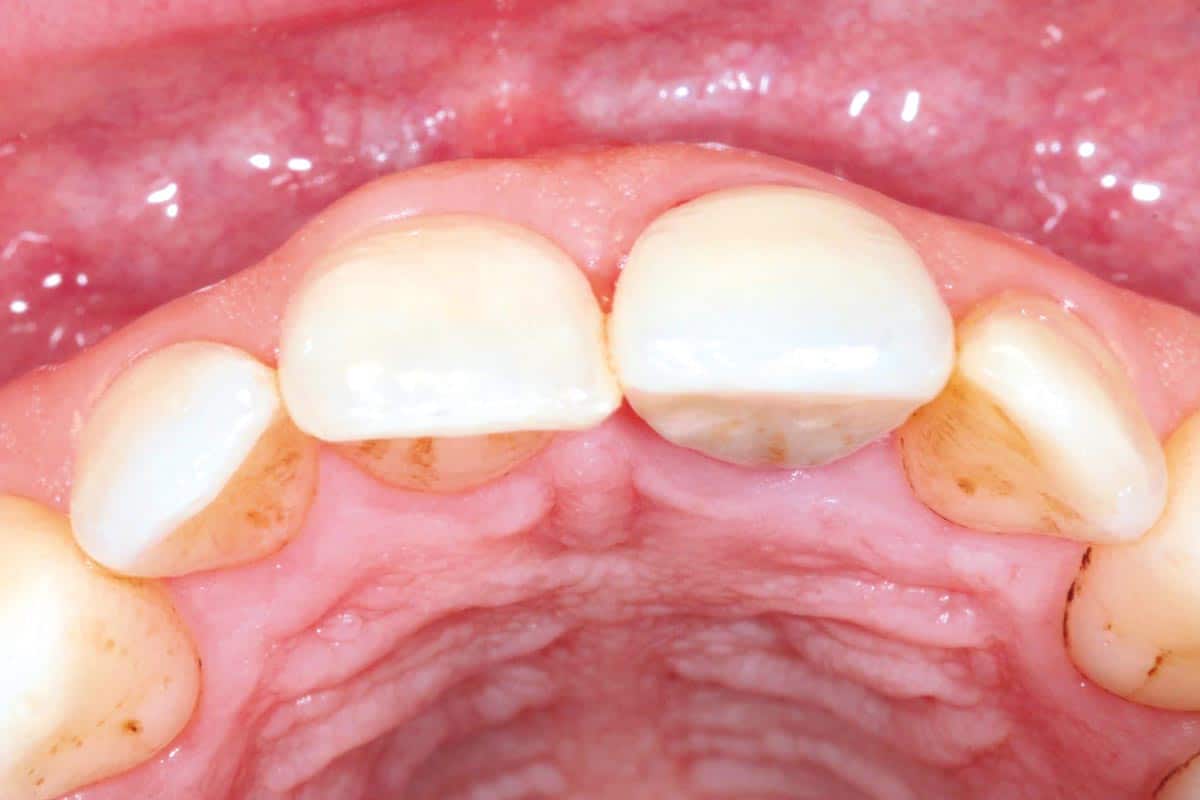
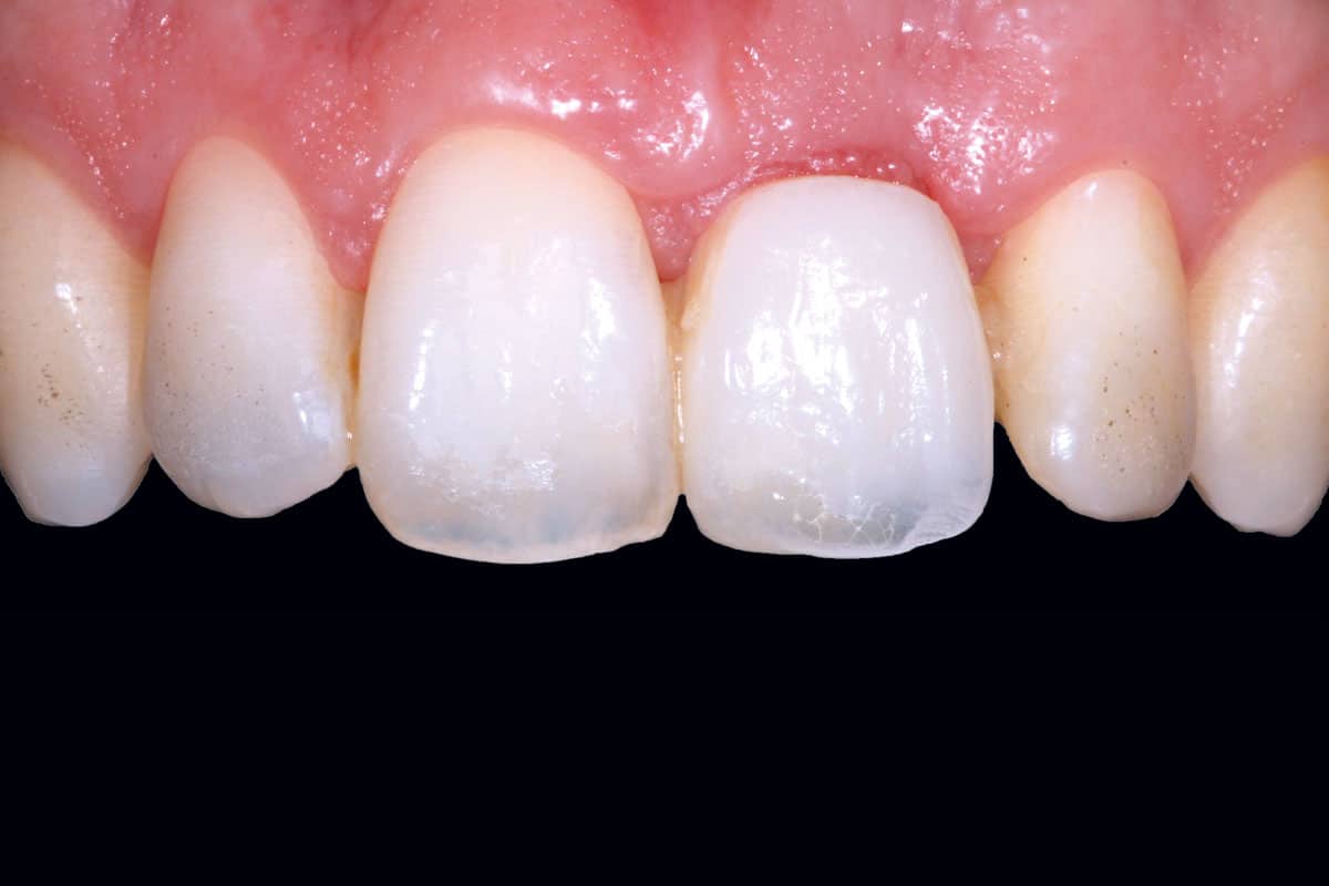
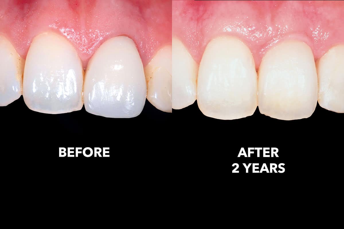
Eight weeks postoperatively, complete soft tissue closure was observed, characterized by healthy keratinized tissue and favorable integration with the surrounding mucosa. The surgical approach demonstrated several clinical advantages, including minimally invasive flap elevation without coronal displacement of the mucogingival junction, preservation of vascular integrity, avoidance of periosteal releasing incisions, and elimination of donor site morbidity due to the omission of autogenous connective tissue harvesting.
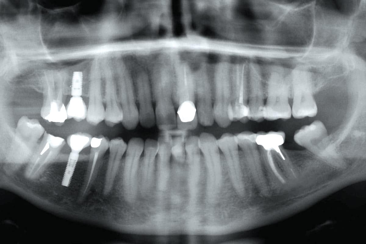
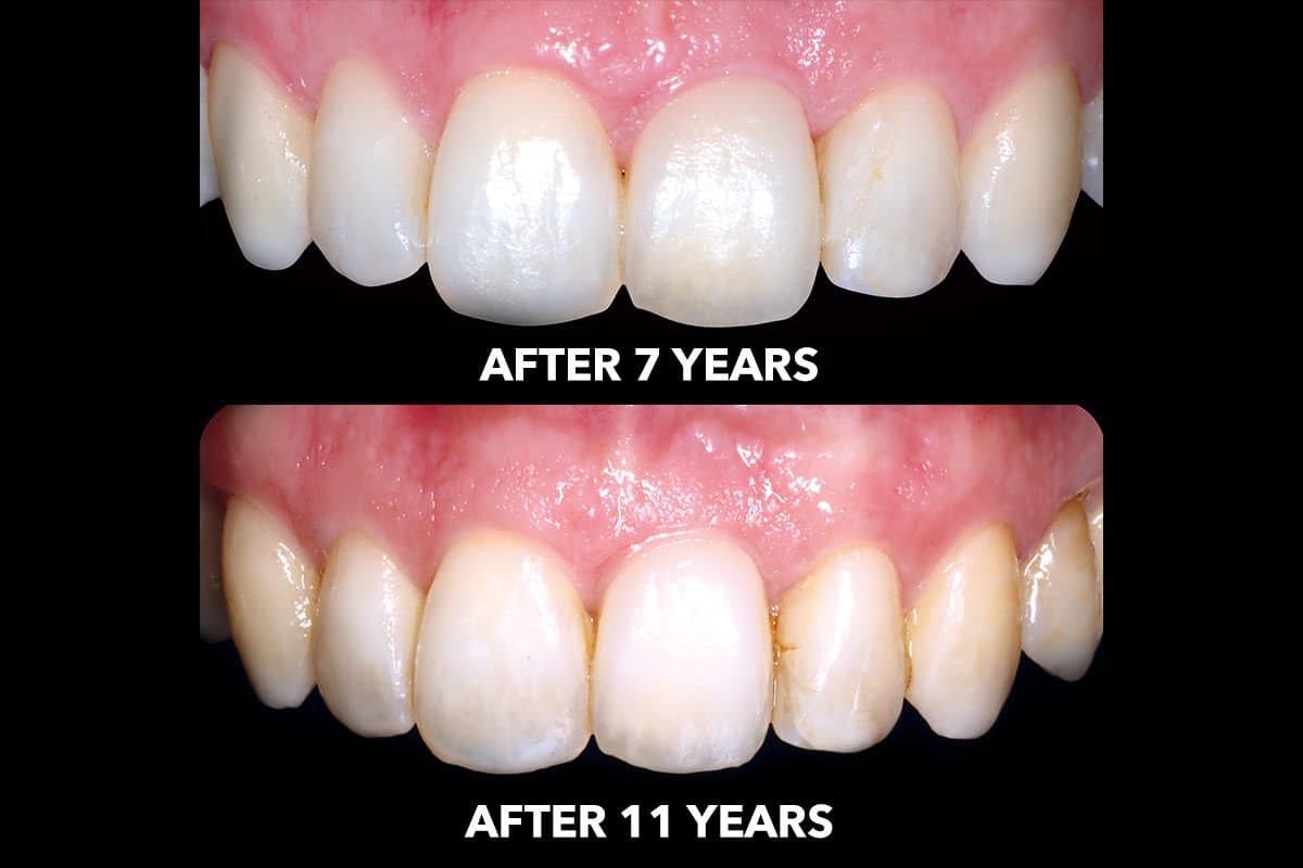
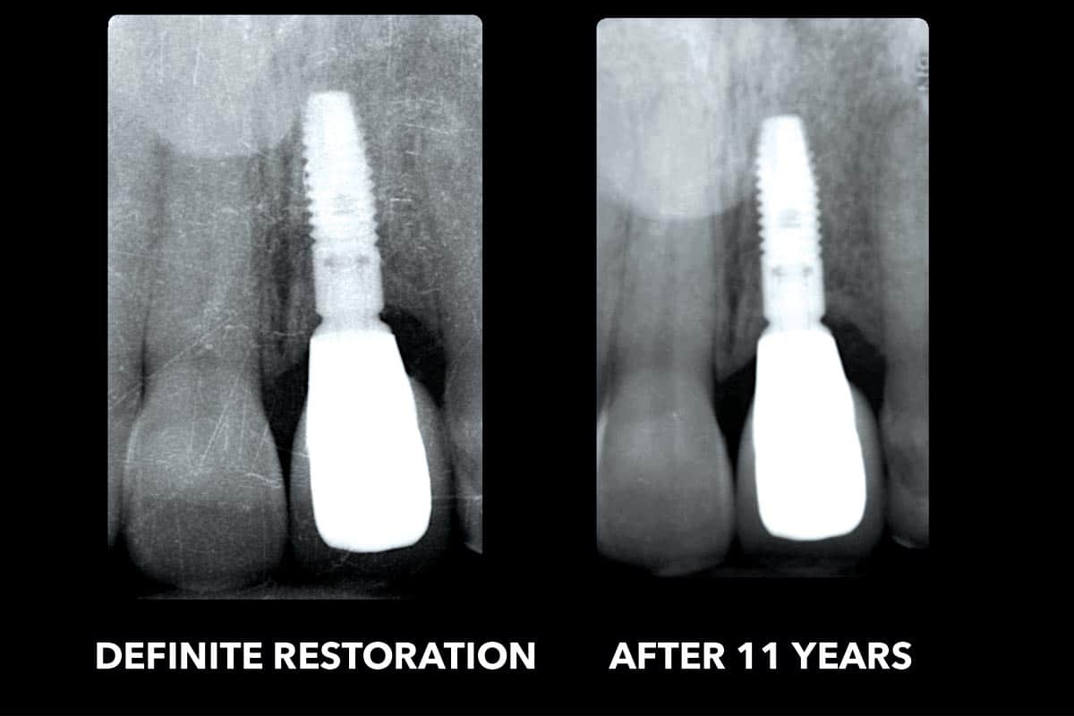
Long-term clinical and radiographic follow-up at seven and eleven years confirmed stable peri-implant hard tissue conditions and maintained peri-implant soft tissue volume. The peri-implant mucosa remained healthy, with no signs of inflammation or recession, underscoring the long-term stability and predictability of this therapeutic approach.
The „Case Of The Month“ highlights every month a clinical case, which distinguished itself by the clinical results or the treatment concept in combination with the applied botiss biomaterials. The selection of the case is based on content relevance and quality of the documentation.


