collacone®
Natural collagen cone for application in extraction sockets

natural collagen
Stabilization of blood clot
Controlled wound healing process
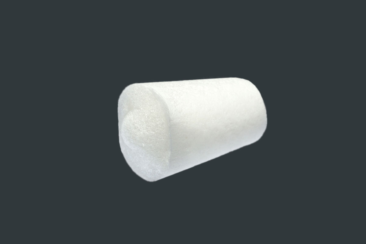
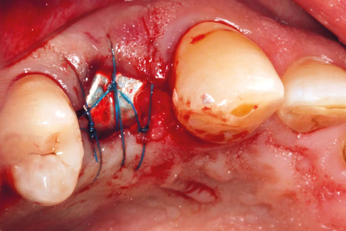
collacone® is a wet-stable and moldable cone made of natural collagen. It was specifically developed and designed for application in fresh extraction sockets, to support the natural healing of the socket. The application of collacone® into the socket supports the stabilization of the formed blood coagulum and helps to control bleeding, while its form-fitted cone shape protects the wound area from food and bacteria 1. collacone® resorbs completely within about 2-4 weeks 2, 3.
BLOOD CLOT PROTECTION AND NATURAL HEMOSTASIS
After tooth removal, the healing of an extraction socket requires the formation and maturation of a blood clot, followed by the infiltration of fibroblasts that replace the coagulum. The spongy structure of collacone® provides an ideal structure for the adhesion of thrombocytes, fibroblasts, and osteoblasts 4, 5. Fine blood vessels grow into and through the cone; as a result, the preliminary tissue formed in the alveolus is supplied with oxygen, nutrients, and the essential signaling molecules, which support its bony regeneration. collacone® application is particularly beneficial in hemostatic compromised patients to prevent postoperative bleeding events 1.


IMPLANTOLOGY, PERIODONTOLOGY AND ORAL AND CMF
collacone® is indicated wherever capillary, venous, small arterial and diffuse seeping bleedings must be stopped, and where conventional means of hemorrhage control are either inadequate or technically difficult and time-consuming. collacone® is used as a hemostatic agent following tooth extractions or harvesting of biopsies, soft tissue and bone transplants.
- Closure of extraction sockets
- Biopsy sites
- Minor oral wounds
- Control and stop of bleeding in extraction sockets or biopsy sites
- Internal sinus lift
Application and shaping
After opening the package, collacone® is taken out with dry sterile instruments, placed and lightly pressed onto the cleaned wound with swabs. collacone® can easily be cut to the necessary size with a sterile pair of scissors.
Rehydration
Generally, dry application of collacone® is recommended, since soaking or moistening the cone prior to implantation may impair its hemostatic properties. At the defect site the fleece rapidly soaks up blood. collacone® maintains its integrity in the presence of blood and during application.
Fixation
At contact with the wet wound surface, the collacone® sticks to the wound and forms a gel like bond with the blood. However, passive fixation by cross- or holding sutures could help keeping the cone in place when applied in extraction sockets.
Exposure
In case of a dehiscence the wound heals without complications by granulation tissue formation. and free contraction. The main problem of an exposure of a collagen membrane is its fast bacterial resorption resulting in loss of the barrier function. As the intention of collacone® is not providing a barrier but supporting wound healing and hemostasis, fast resorption in case of open healing is not a problem.
- Resorption within two to four weeks
- Stabilization of blood clot and efficient local hemostasis
- Maintains integrity in the presence of blood and during application
- Wound protection
- Highly porous structure for ingrowth of vessels and cells
- Controlled wound healing process
- Natural collagen cone

Distribution
With our international network of distribution partners, we are near you in over 100 countries worldwide. In addition to our 360° productportfolio, we offer service, scientific advice and exchange, training and events directly on site from a single source.
Find a distribution partner near you:
SPECIFIC FACTS
CASES


Science
- Study


Education


Edutainment
- RECORDED-WEBINAR

 Dr. Hassan MaghairehUnited Kingdom


