CLINICAL CASE
Dr. Lucas Werutsky
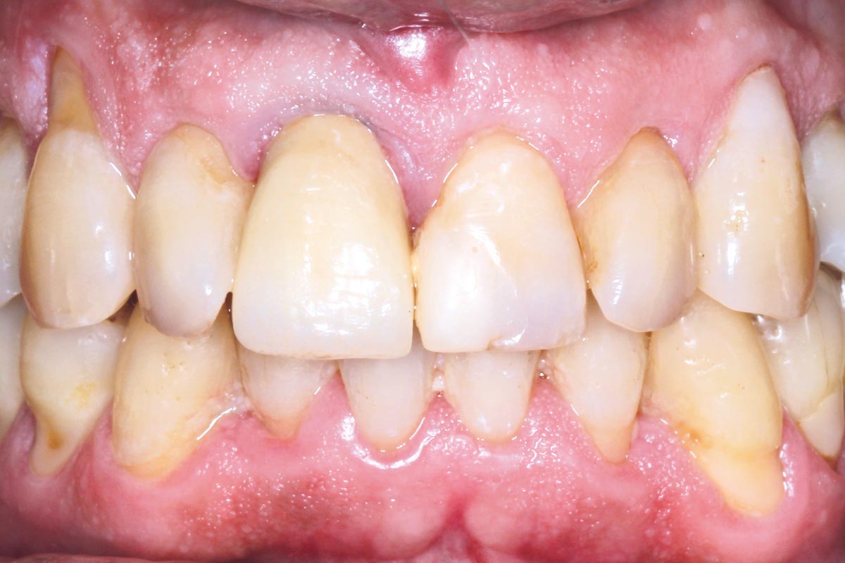
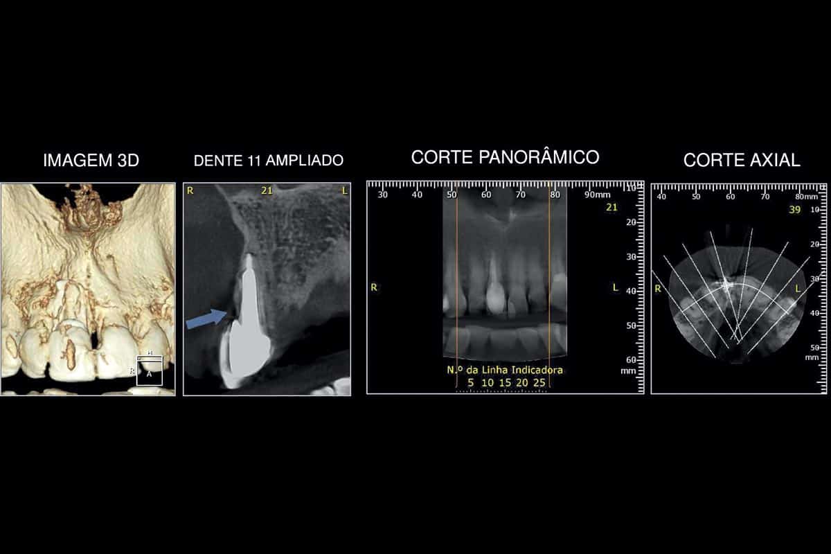
In this clinical case, a patient presented a cracked dental root in the incisor region, specifically on tooth 11, rendering prosthetic rehabilitation of the natural tooth highly challenging. In addition, the patient exhibited gingival recession at the affected site. The tomographic images confirmed the presence of an oblicual root fracture.
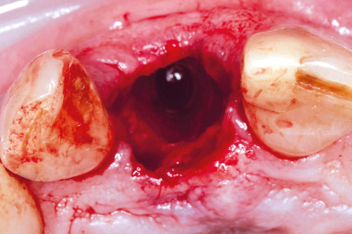
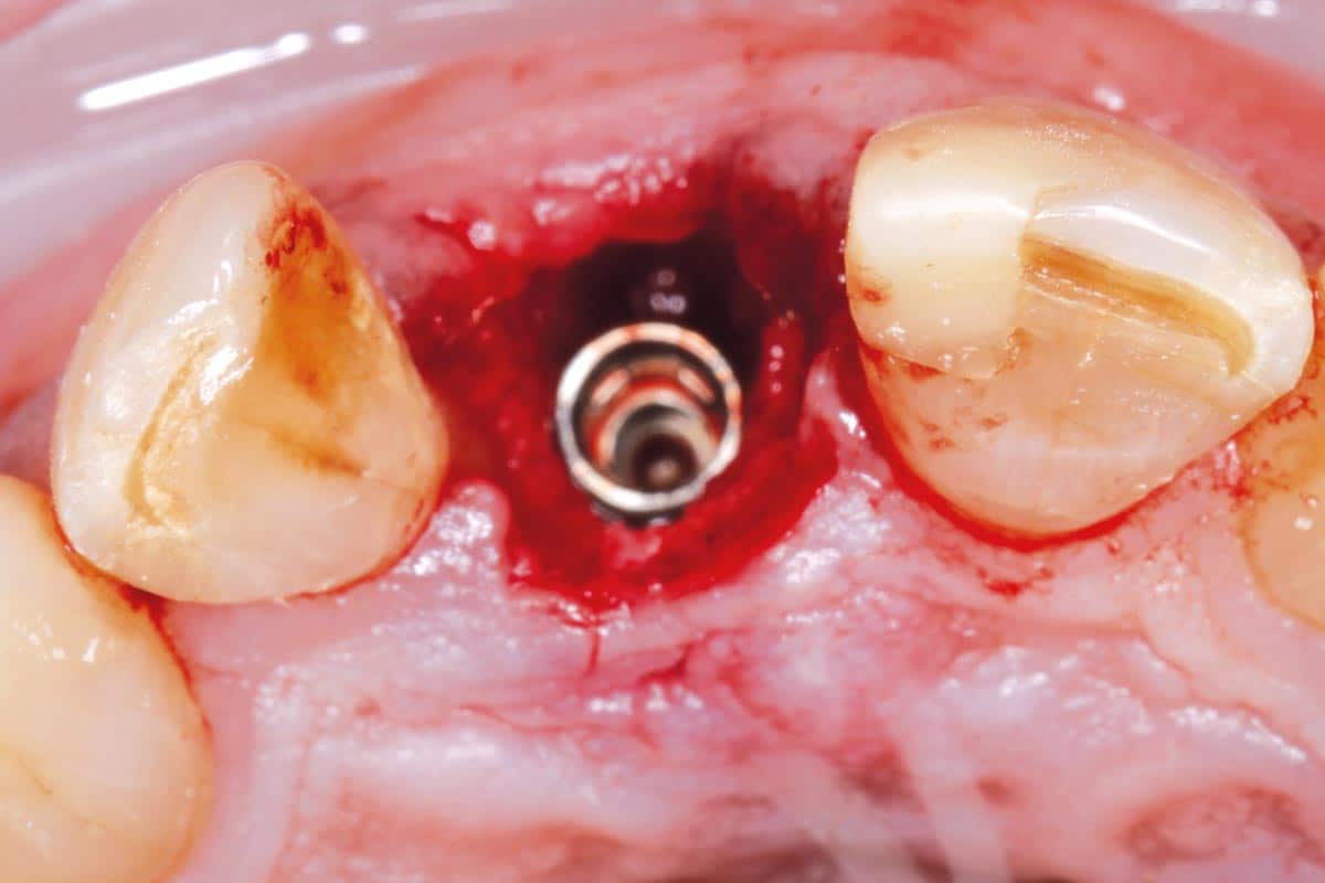
Based on the clinical and radiographic findings, the treatment plan consisted of an atraumatic tooth extraction followed by an immediate implant placement in order to preserve hard and soft tissue architecture and optimize the aesthetic outcome.
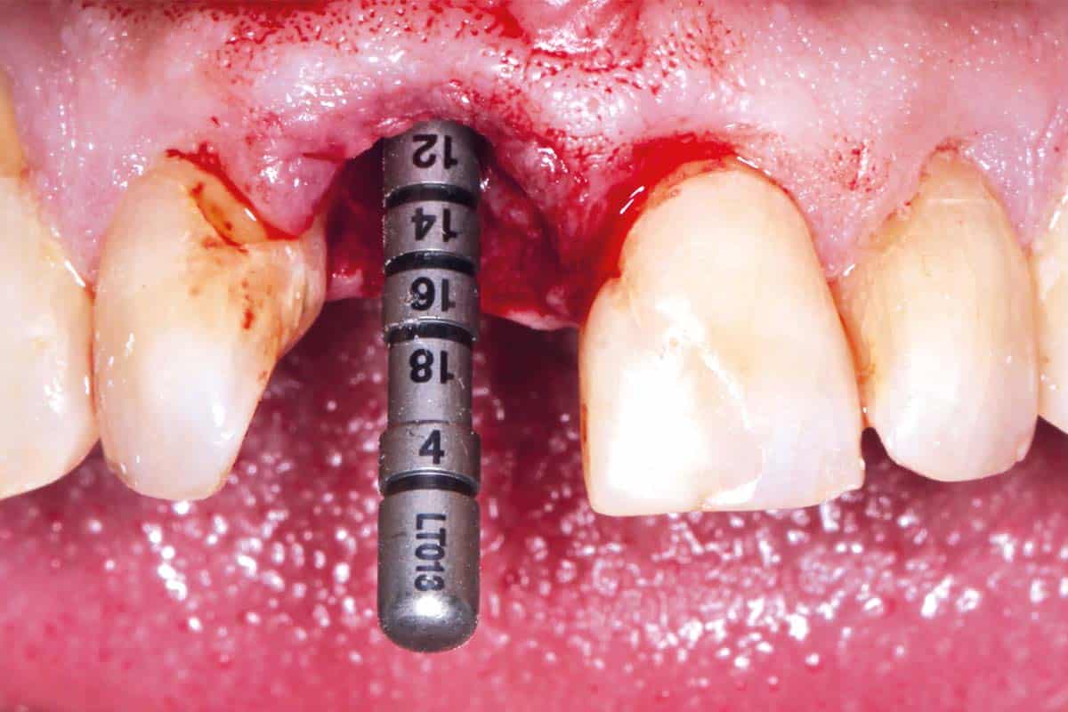
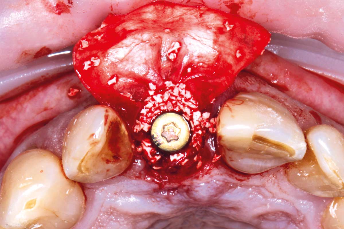
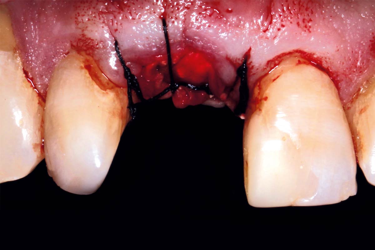
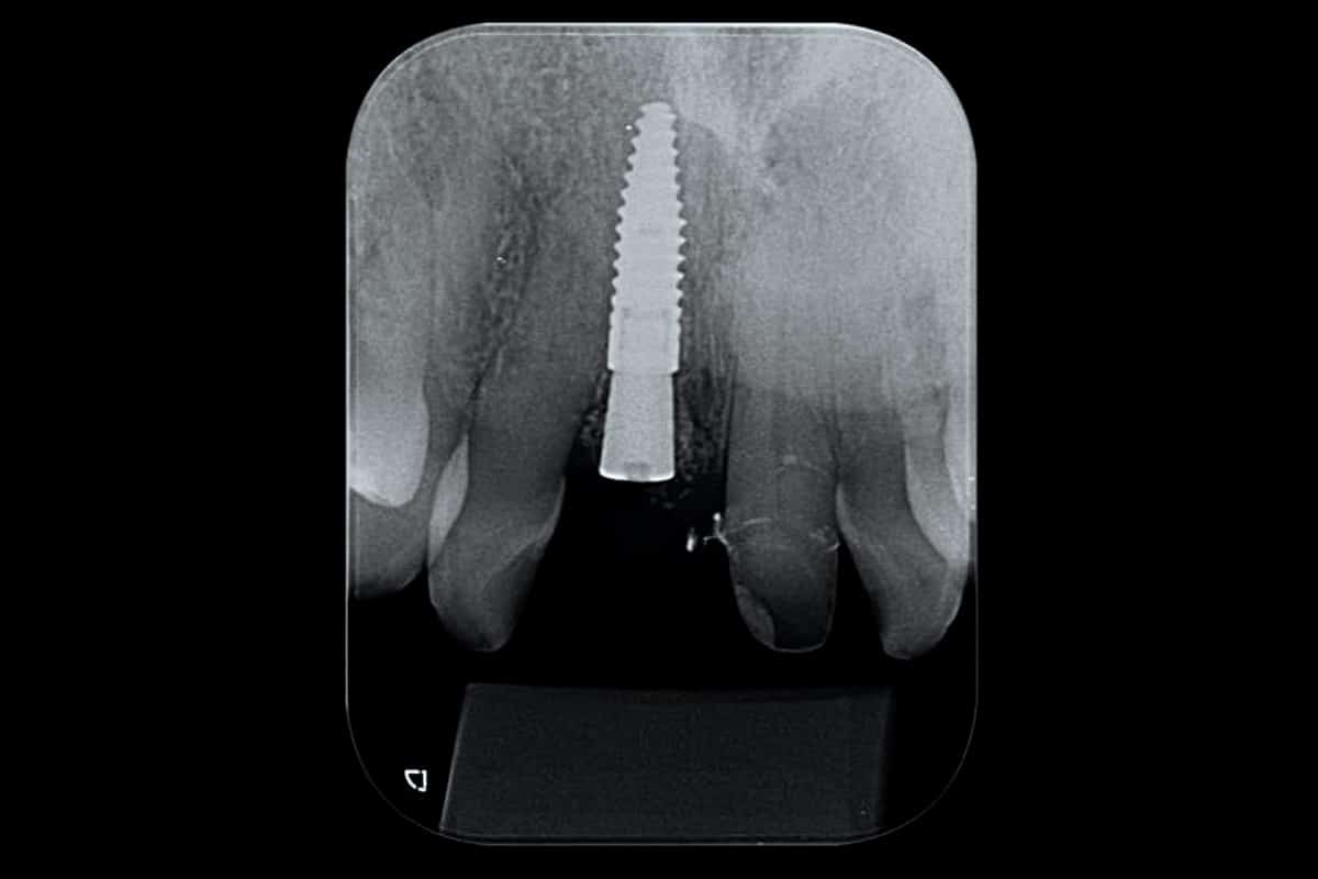
Following extraction, an implant was placed immediately into the extraction socket. A buccal gap was present and was managed through ridge reconstruction using cerabone®, providing a stable and osteoconductive scaffold for new bone formation. The grafted area was covered with a Jason® membrane to stabilize the grafting material and support guided bone regeneration (GBR). Wound closure was achieved using sutures.
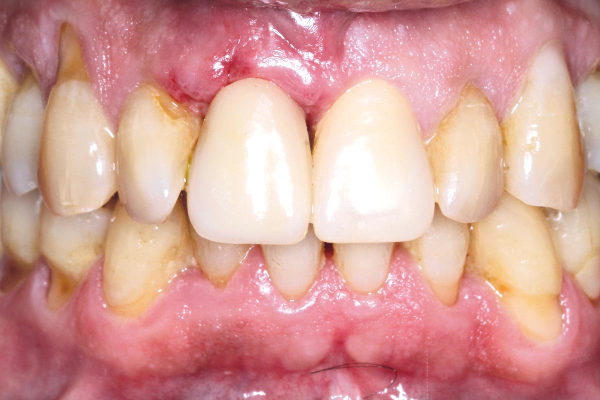
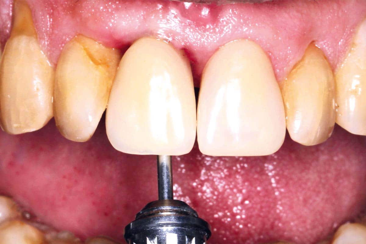
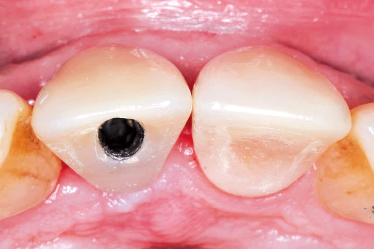
Tooth 21 was prepared for a full-coverage crown, and bonded provisional crowns were placed on both tooth 11 and tooth 21 to maintain aesthetics and soft tissue support during healing. After 30 days, a provisional crown was placed on implant site 11 to refine and condition the peri-implant soft tissue contour.
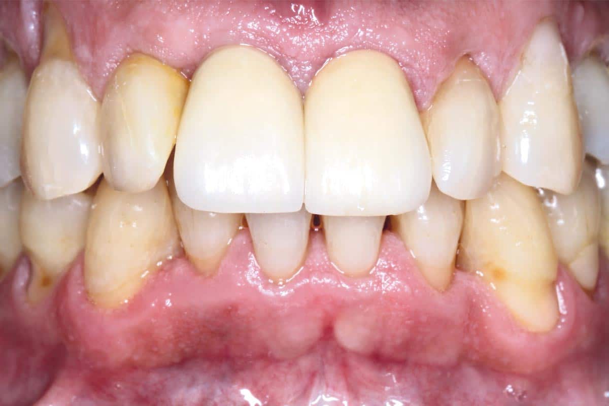
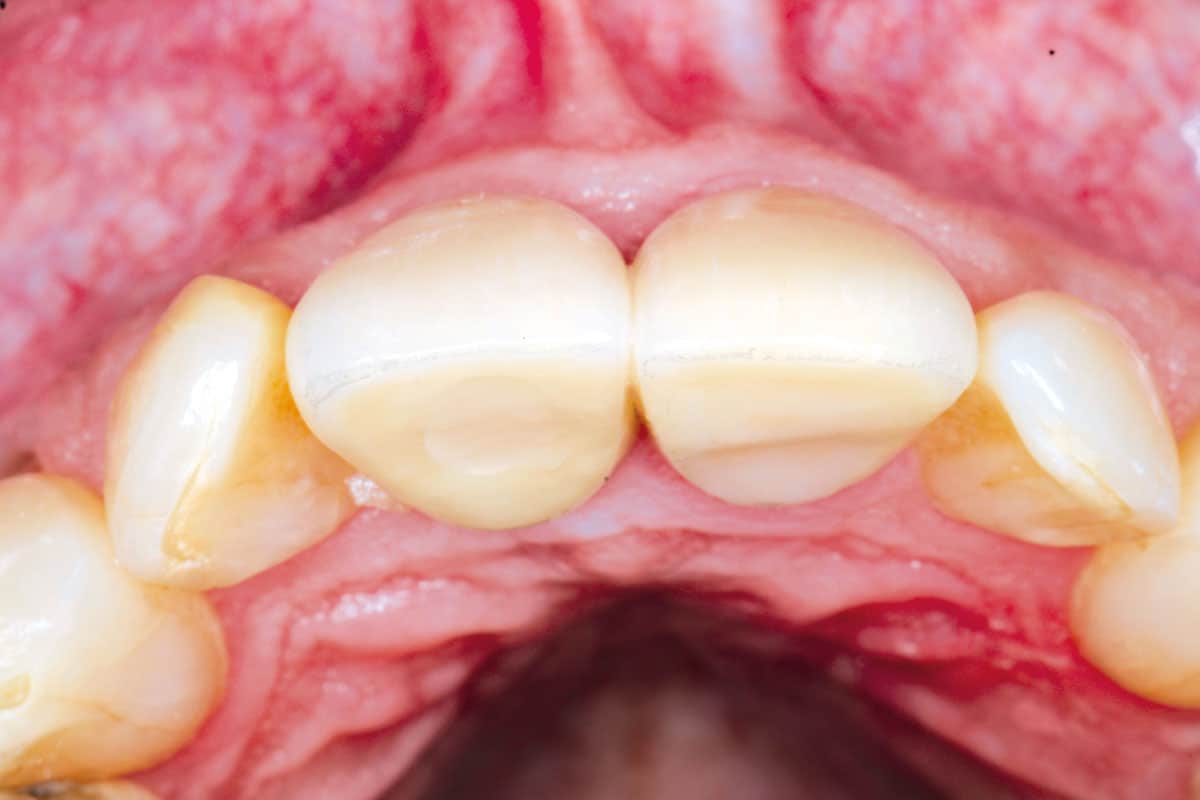
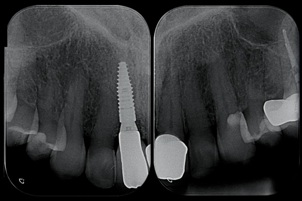
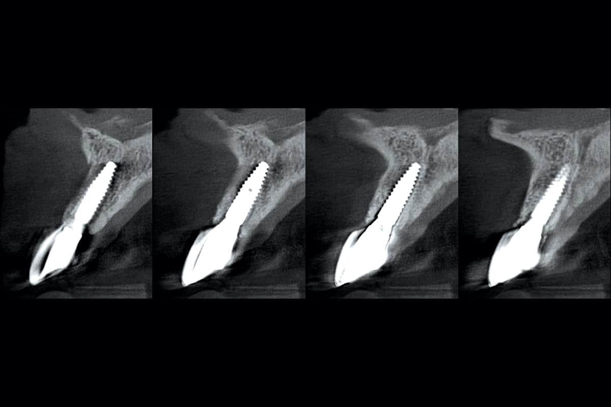
The final prosthetic rehabilitation consisted of ceramic crowns placed on implant site 11 and tooth 21. A one-year post-restoration CBCT evaluation demonstrated a stable clinical and radiographic situation with satisfactory graft integration. These findings highlight the predictable performance of cerabone® in combination with Jason® membrane and confirm the reliability of this approach for long-term implant success in the aesthetic zone.