CLINICAL CASE
Dr. Robert Williams
Clinical Situation
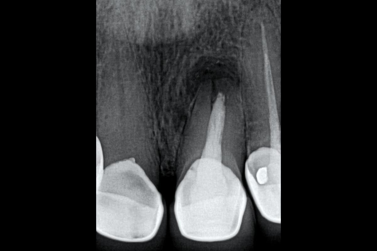
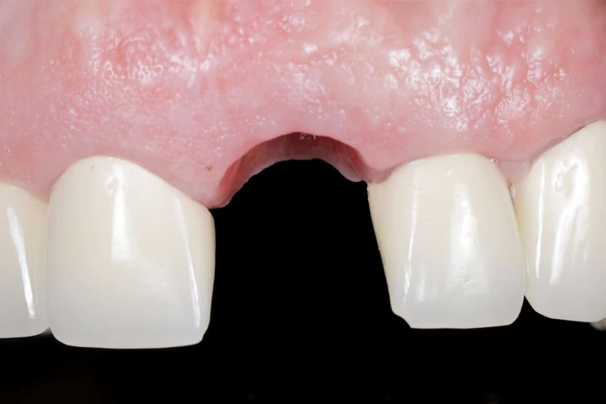
Preoperative radiographic assessment confirmed thin/missing buccal bone and a high risk of resorption following conventional extraction.
In the anterior maxilla, preservation of the facial bone and gingival architecture is critical to achieve predictable aesthetic outcomes.
To minimize post-extraction ridge remodeling and maintain the natural contour of the alveolar ridge, an immediate implant placement protocol combined with the NOVAMag® SHIELD technique was selected.
Surgical Procedure
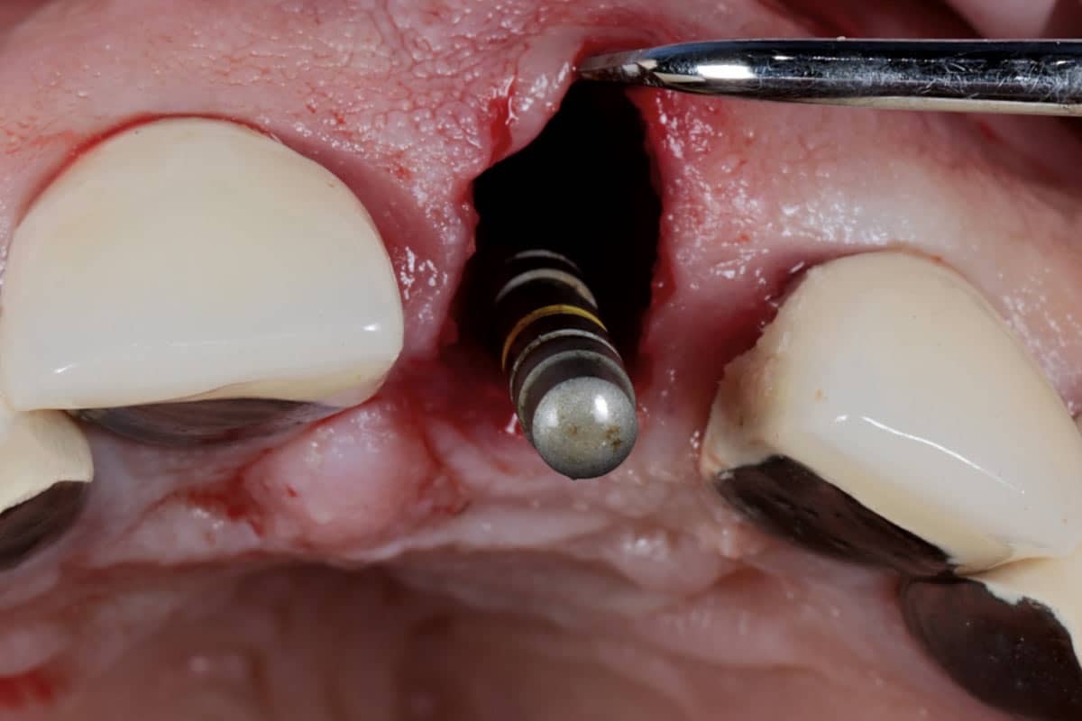
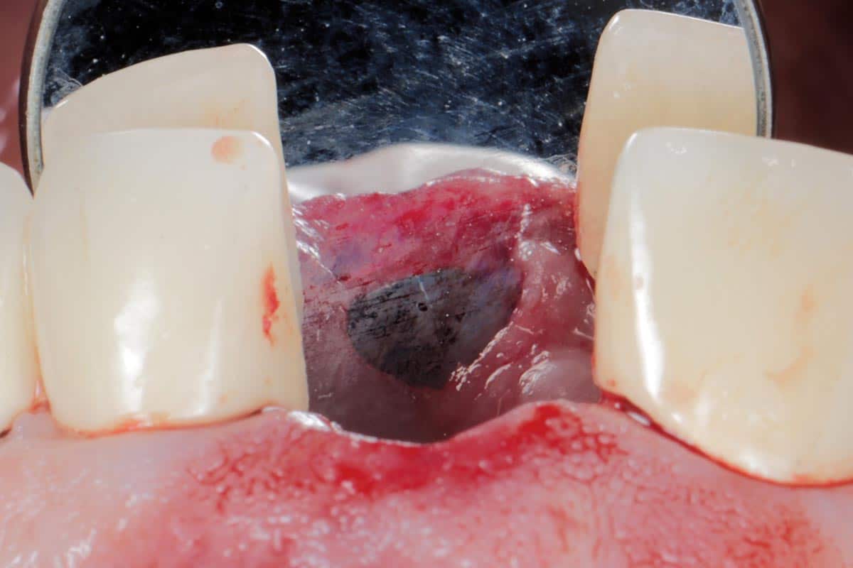
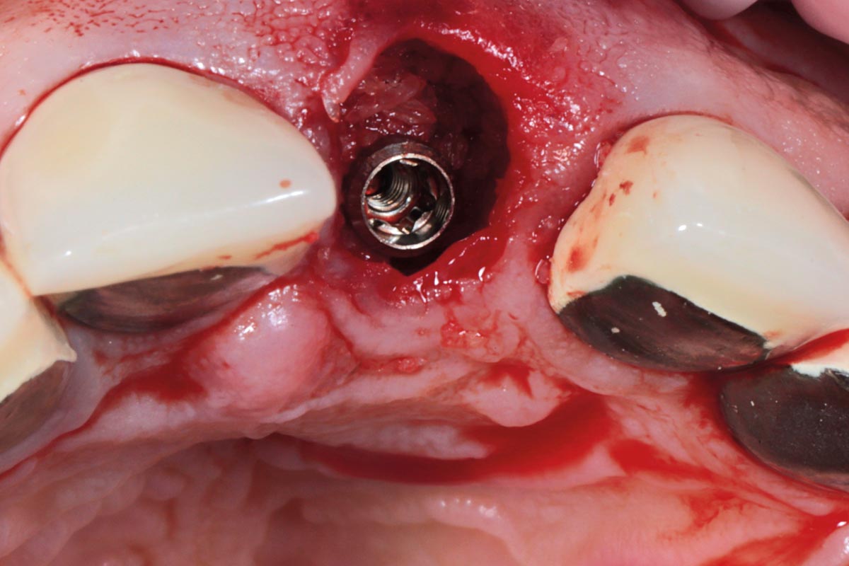
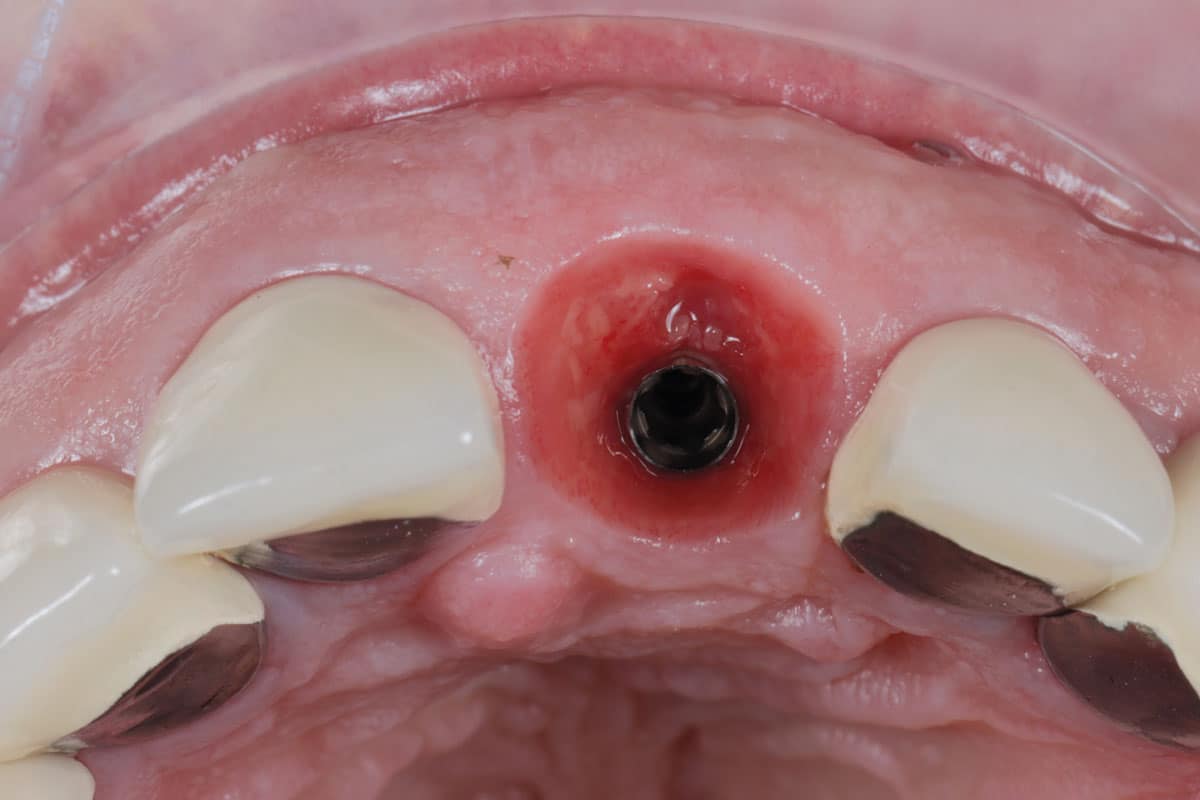
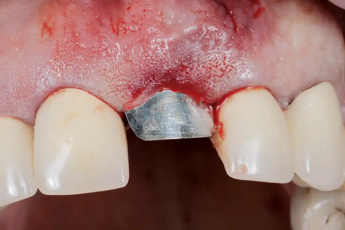
The compromised maxillary central incisor was extracted. The implant was placed in the correct three-dimensional position within the socket.
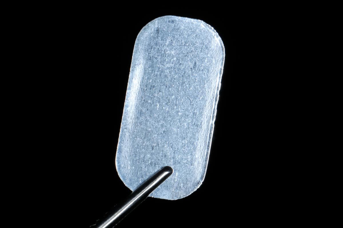
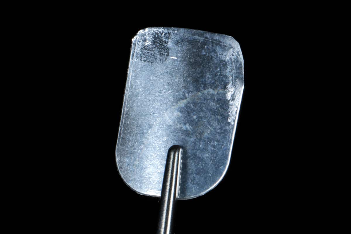
To reconstruct and support the buccal bone plate, a NOVAMag® SHIELD was used. The magnesium shield was carefully shaped and trimmed to match the anatomical contour of the labial socket wall.
The shield was then positioned on the labial wall of the socket, between bone and periosteum, to maintain the ridge contour. The jumping gap was grafted with maxgraft® particles to ensure implant stability. Finally, an immediate temporary crown was fabricated and placed.
Healing and Follow-Up
During the healing period, the NOVAMag® SHIELD provided structural support for the buccal bone and facilitated guided bone regeneration in the facial aspect of the implant.
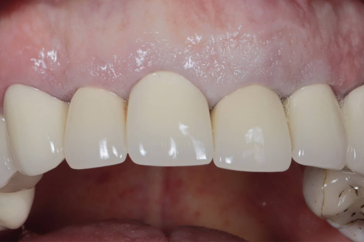
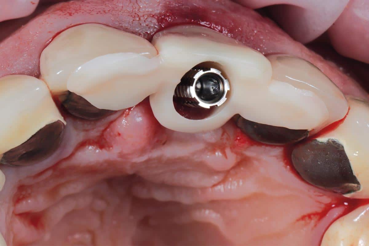
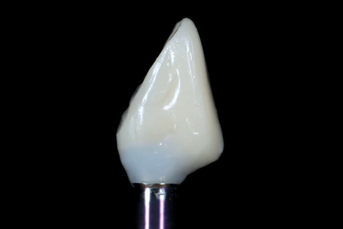
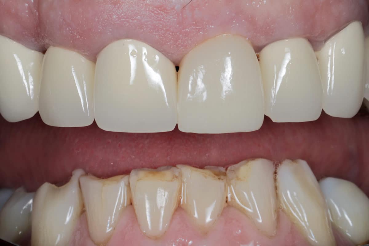
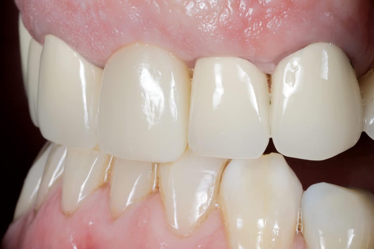
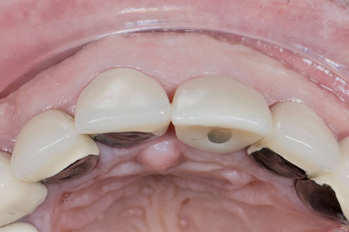
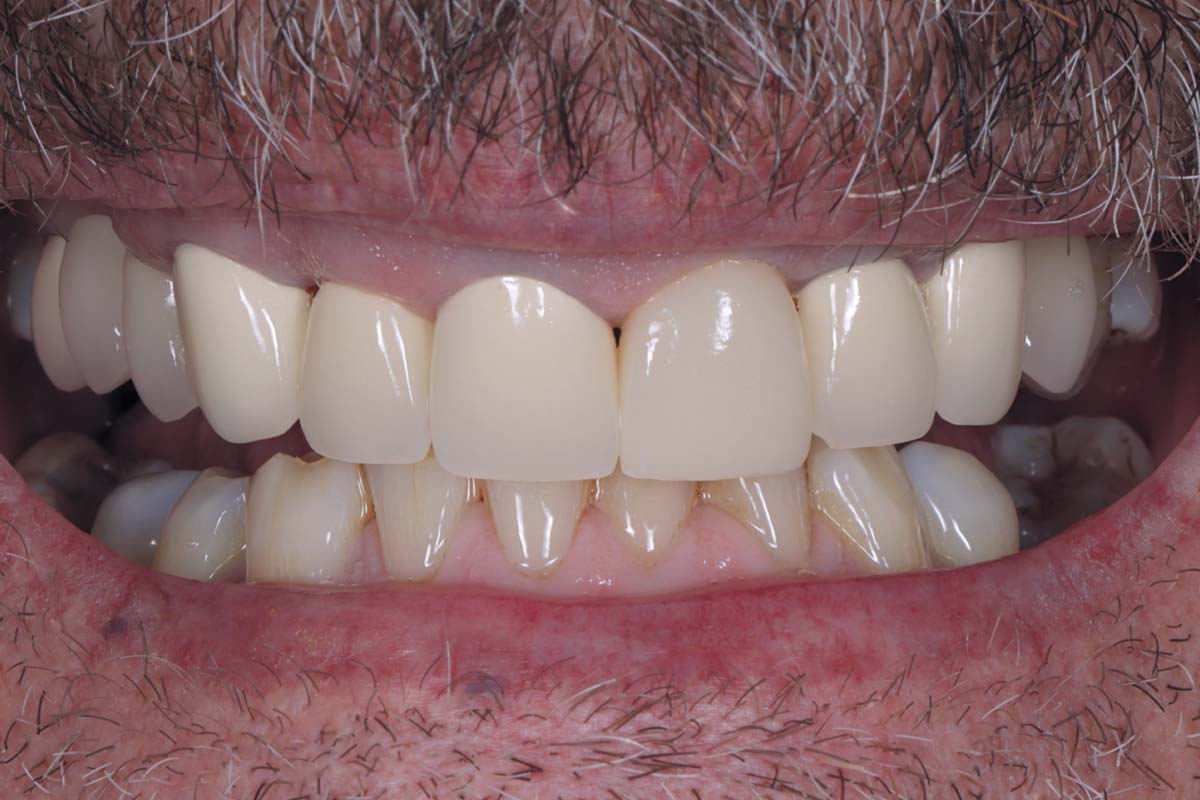
After 10 weeks of healing and successful osseointegration of the implant, the temporary crown was removed and replaced with a definitive prosthetic restoration designed to replicate the natural morphology of the maxillary central incisor.
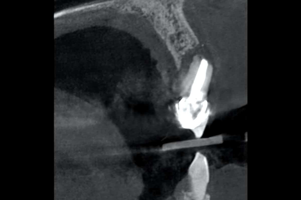
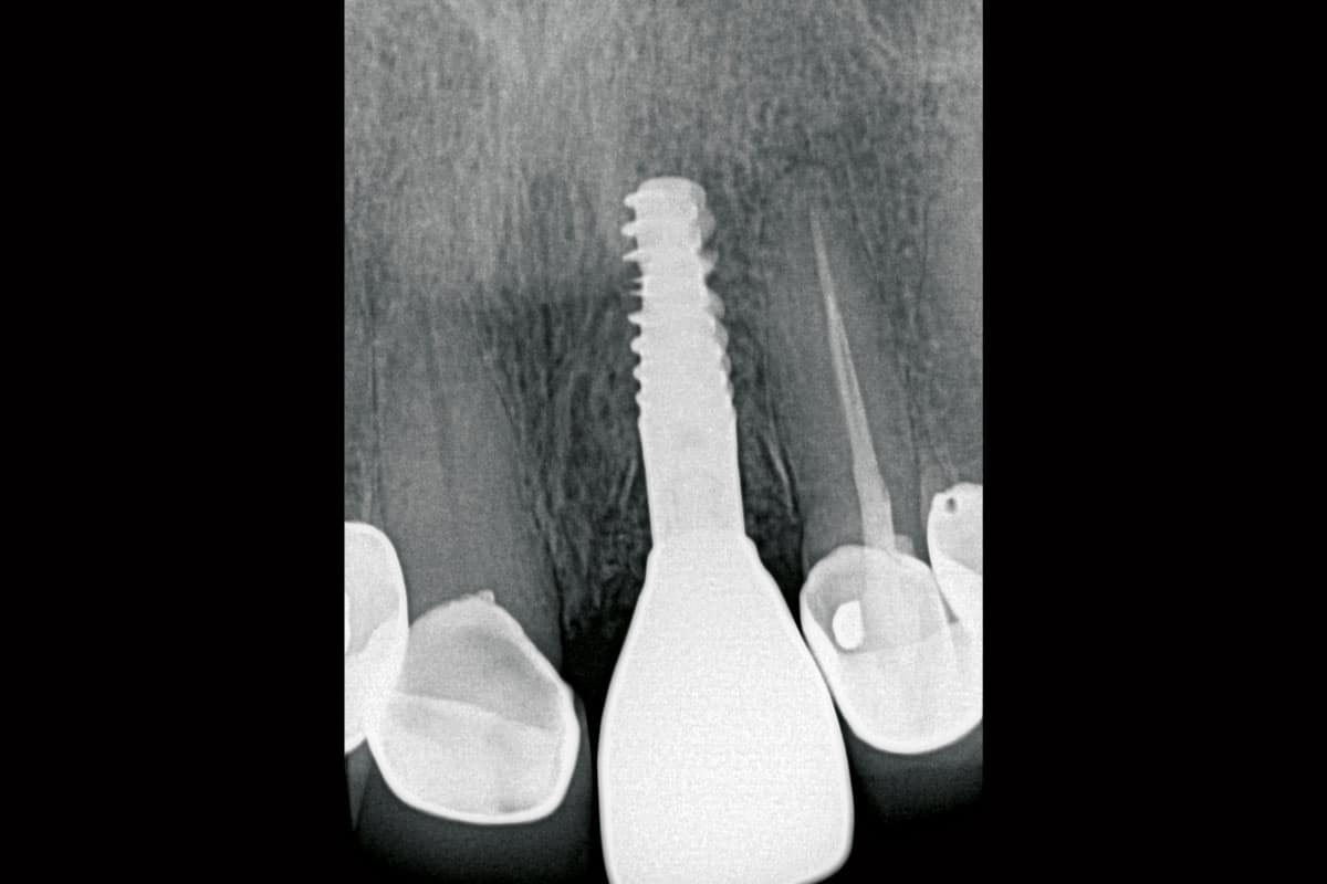
A postoperative radiograph confirmed stable implant positioning and satisfactory peri-implant bone conditions, demonstrating successful hard and soft tissue preservation following the regenerative approach.
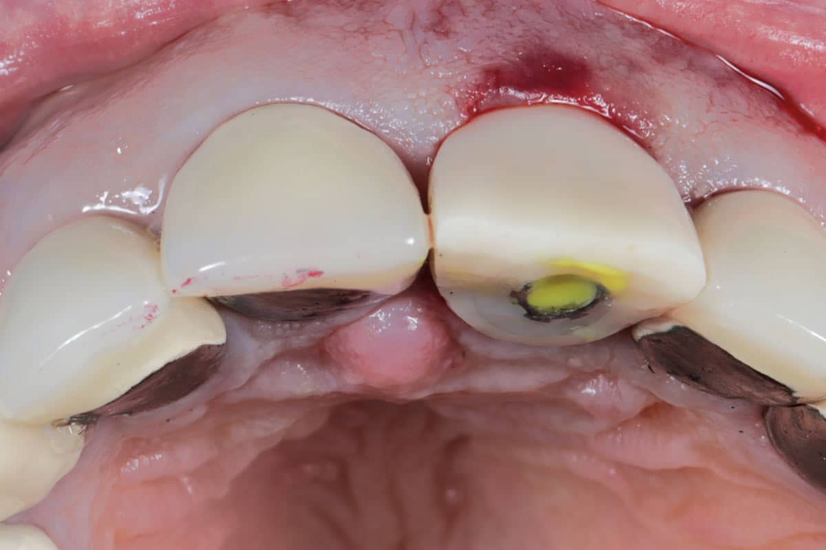
The final outcome showed stable peri-implant tissues and predictable aesthetic results in the anterior maxilla.