CLINICAL CASE
Dr. Ketkee P Asnani
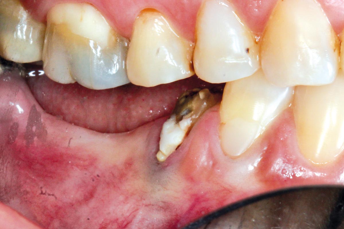
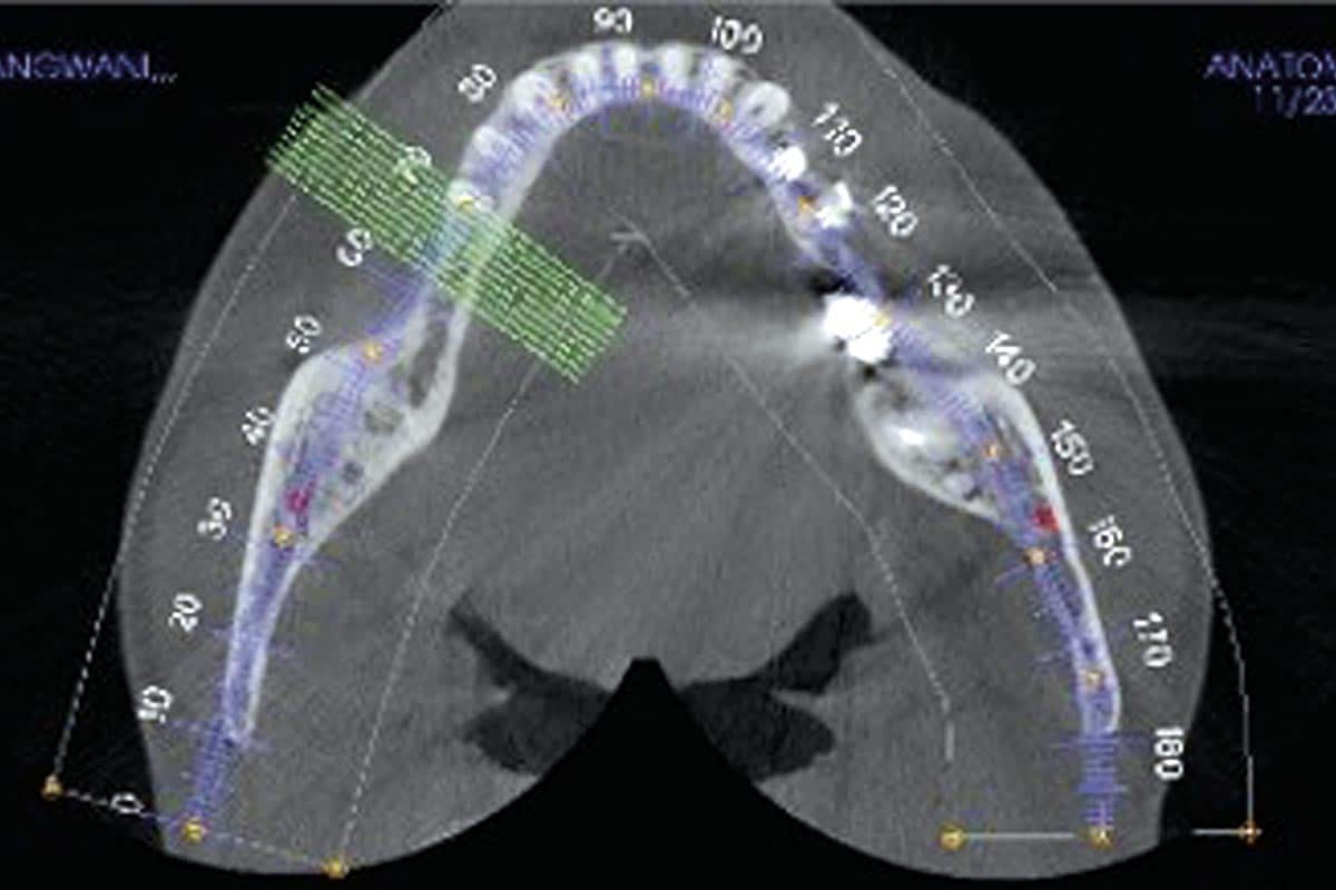
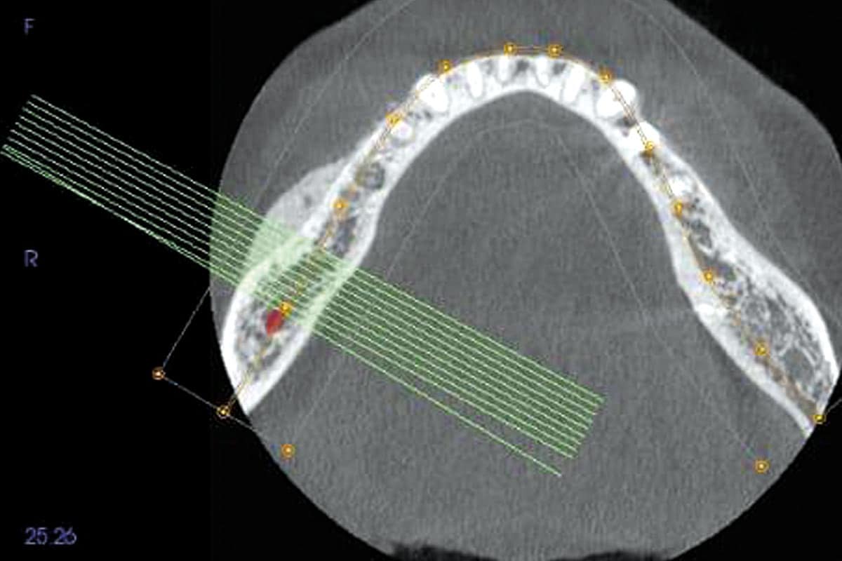
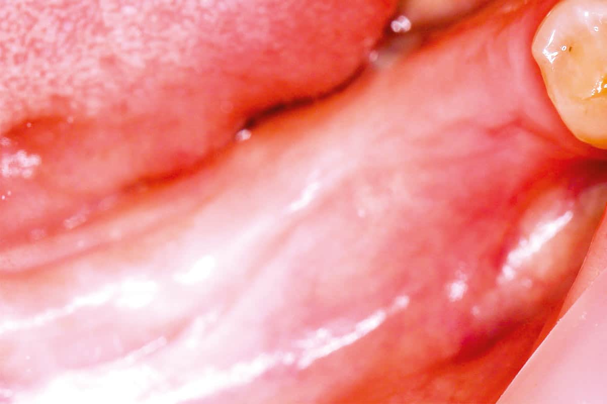
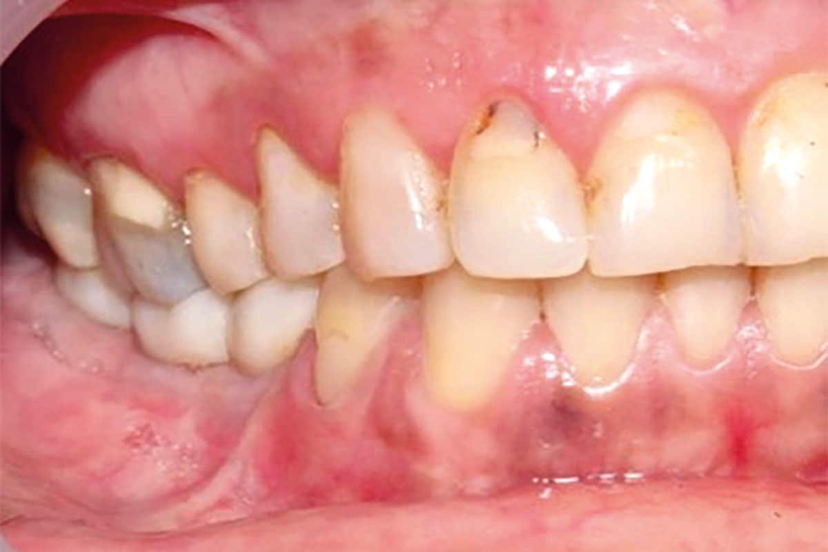
A 45-year-old healthy female presented with a dislodged bridge in the lower right posterior mandible. Clinical examination revealed compromised abutment health, making preservation of the existing prosthetic solution unfavorable. Considering the patient’s age, excellent systemic health, and desire for a long-term fixed rehabilitation, a treatment plan involving horizontal ridge reconstruction followed by implant placement was established. Pre-operative CBCT analysis demonstrated significant horizontal bone loss in the affected region. The treatment objective was to restore ridge width using a combination graft composed of autogenous bone and cerabone®.
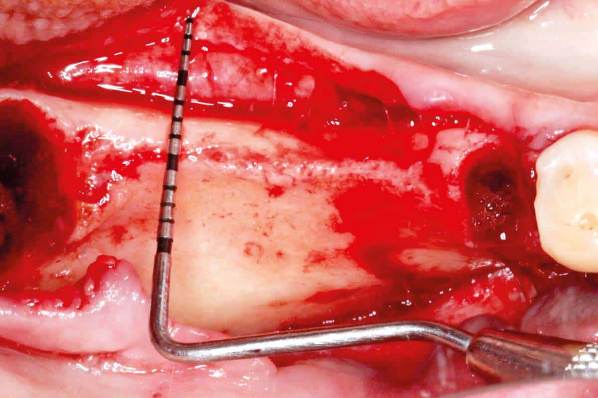
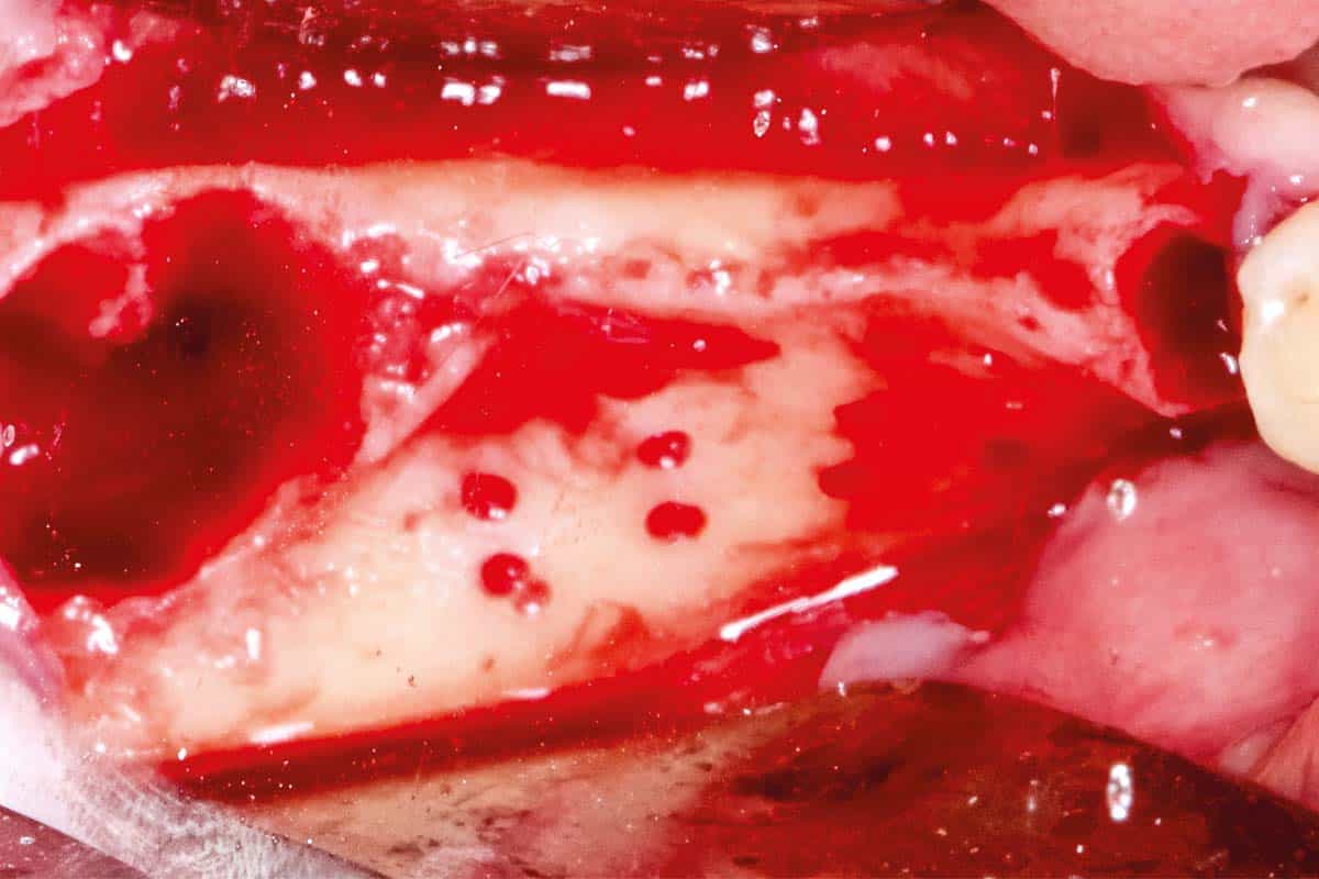
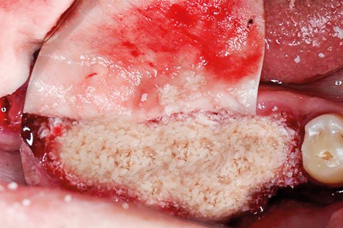
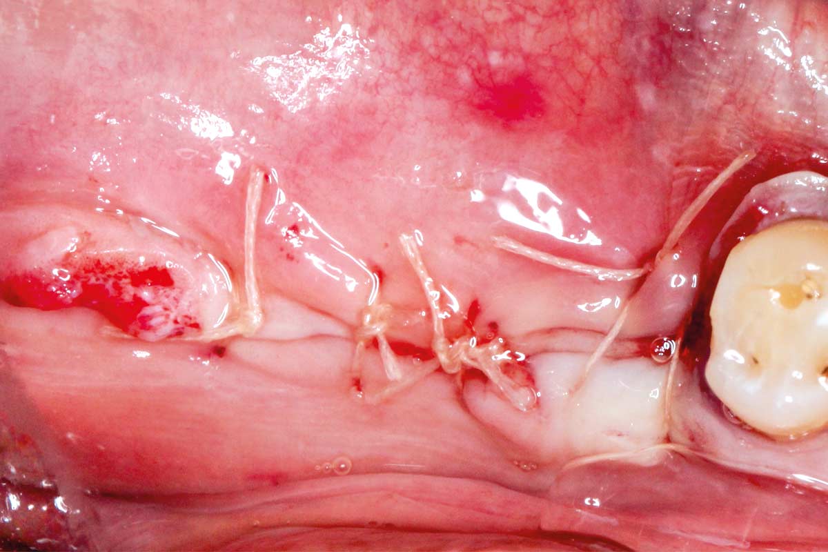
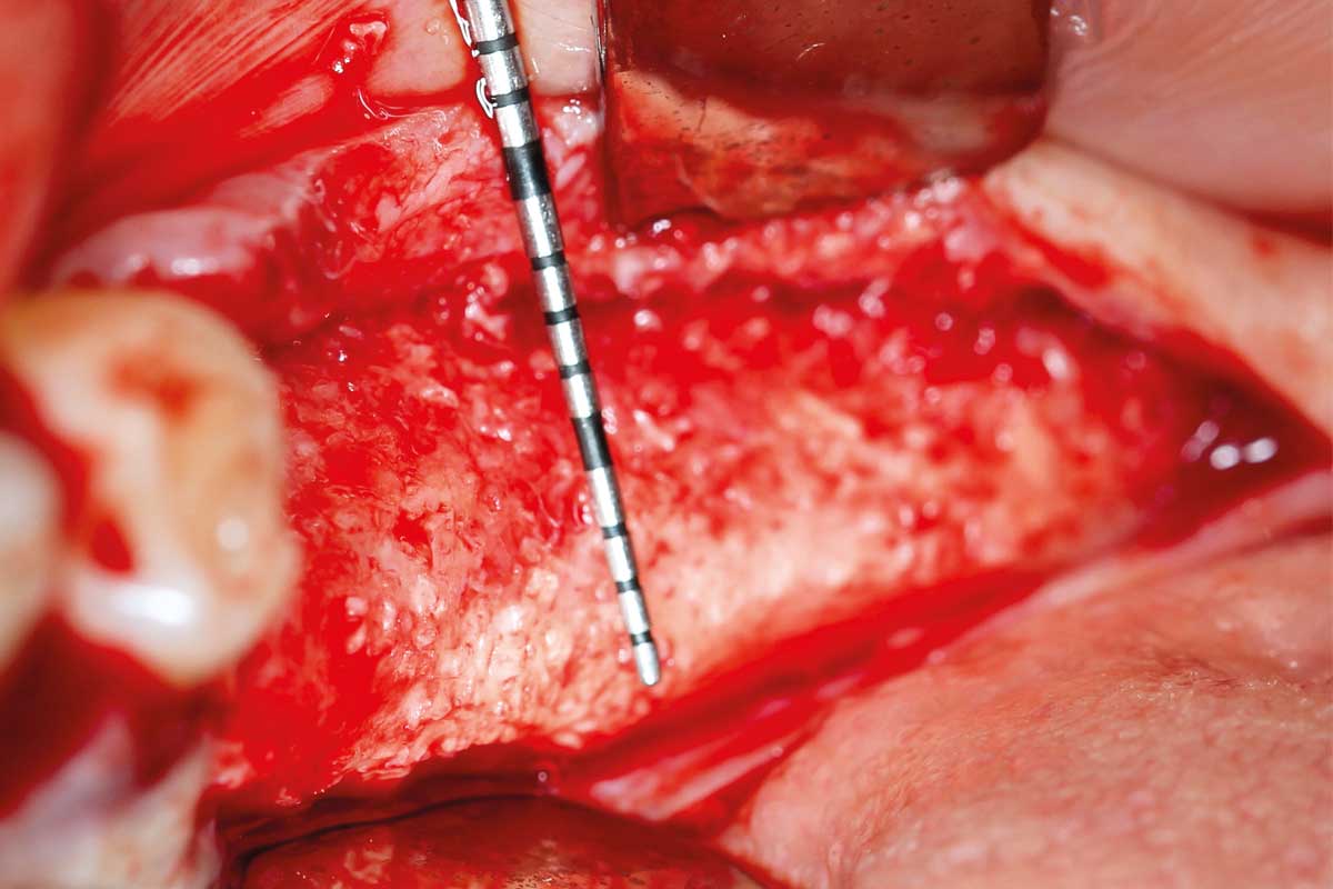
Following flap reflection, a severely atrophied alveolar ridge was observed, with approximately 2.5 mm of crestal width. Decortication of the recipient site was performed to open the marrow spaces and enhance vascularization, thereby promoting graft integration. The grafting material consisted of autogenous bone harvested with a bone scraper combined with cerabone®, which ensures long-term volume stability while serving as an osteoconductive scaffold to support new bone formation. A resorbable collagen membrane was positioned and first stabilized lingually with tacks. The horizontal ridge was then augmented using the particulate graft mixture. The membrane was tightly secured and pinned, ensuring immobilization of the graft material and creating a stable environment for guided bone regeneration. This approach enabled extended horizontal augmentation while maintaining graft stability. Primary wound closure was achieved using a combination of mattress and single interrupted sutures. Immediate post-operative CBCT imaging confirmed a well-compacted graft with securely placed fixation tacks.
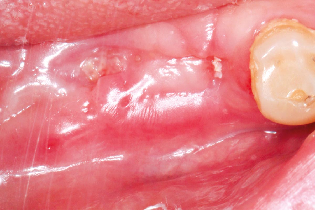
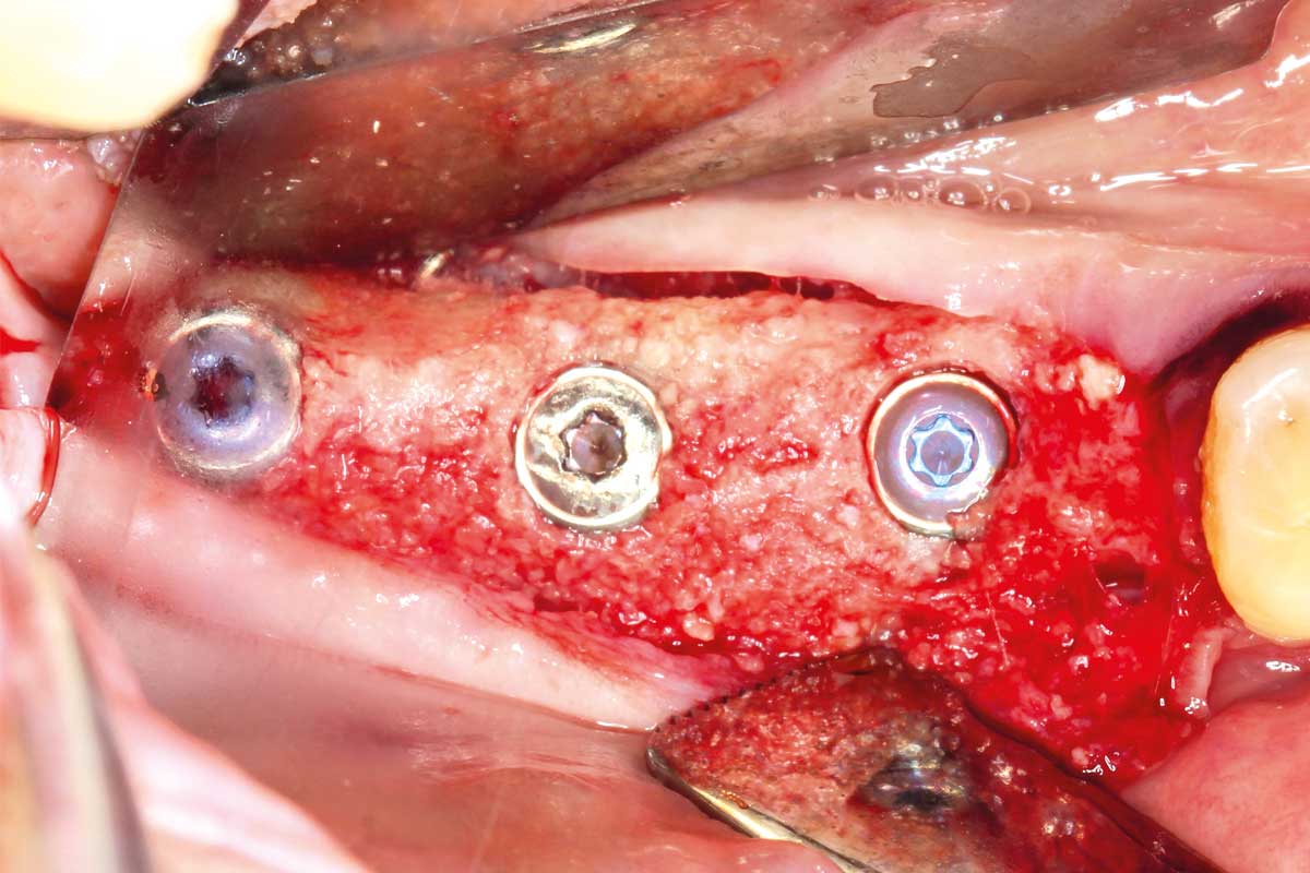
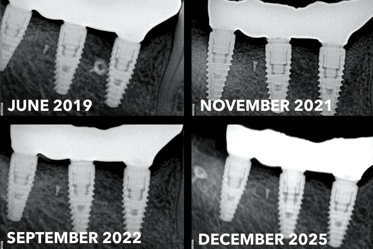
A healing period of at least six months was respected to allow bone formation and maturation. At eight months, flap reflection revealed a significant gain in ridge width and healthy keratinized tissue. Sufficient regenerated bone volume permitted the placement of three implants using a submerged healing protocol.
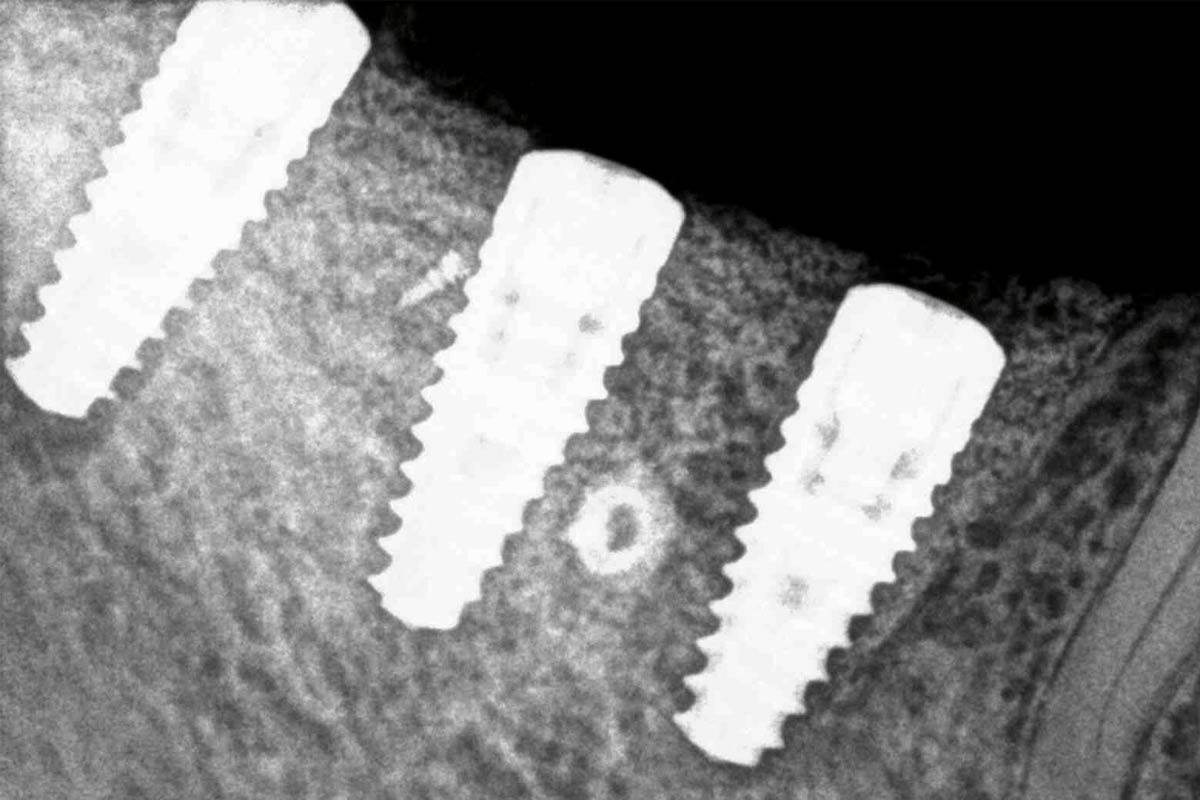
Long-term follow-up at 6.5 years demonstrated stable peri-implant bone levels and healthy surrounding soft tissue, confirming the predictability and durability of the horizontal augmentation procedure using a combined autogenous bone and xenograft approach.