

CLINICAL CASE
Dr. Tolga Genç
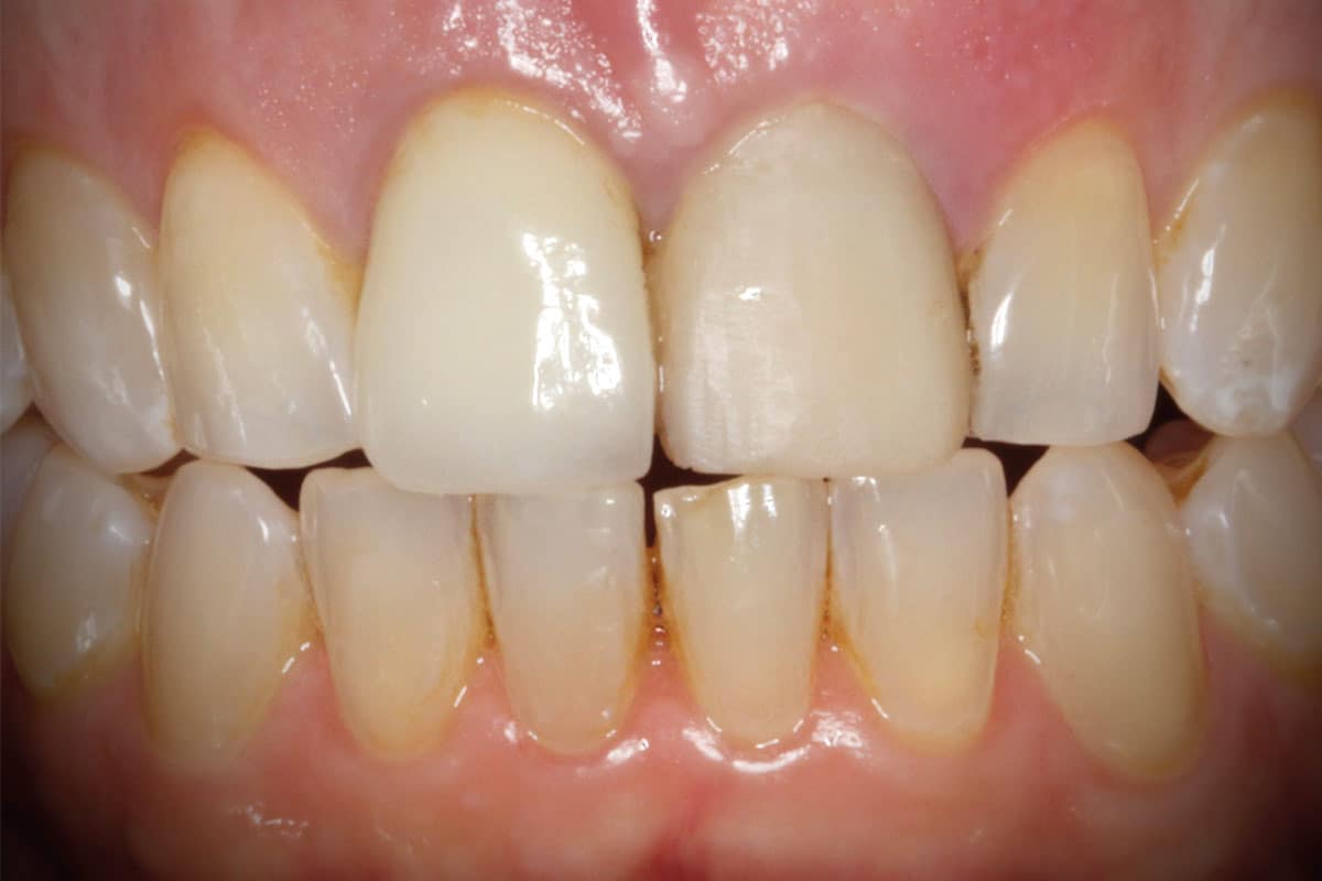
A 60-year-old female patient presented with pain and swelling associated with the maxillary right central incisor 11. Her medical history revealed that the tooth had undergone root canal treatment and apicoectomy approximately 20 years earlier. Two weeks before presentation, the tooth had sustained trauma during intubation under general anesthesia.
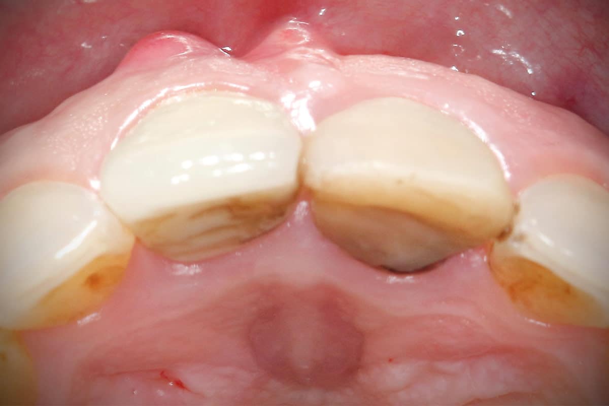
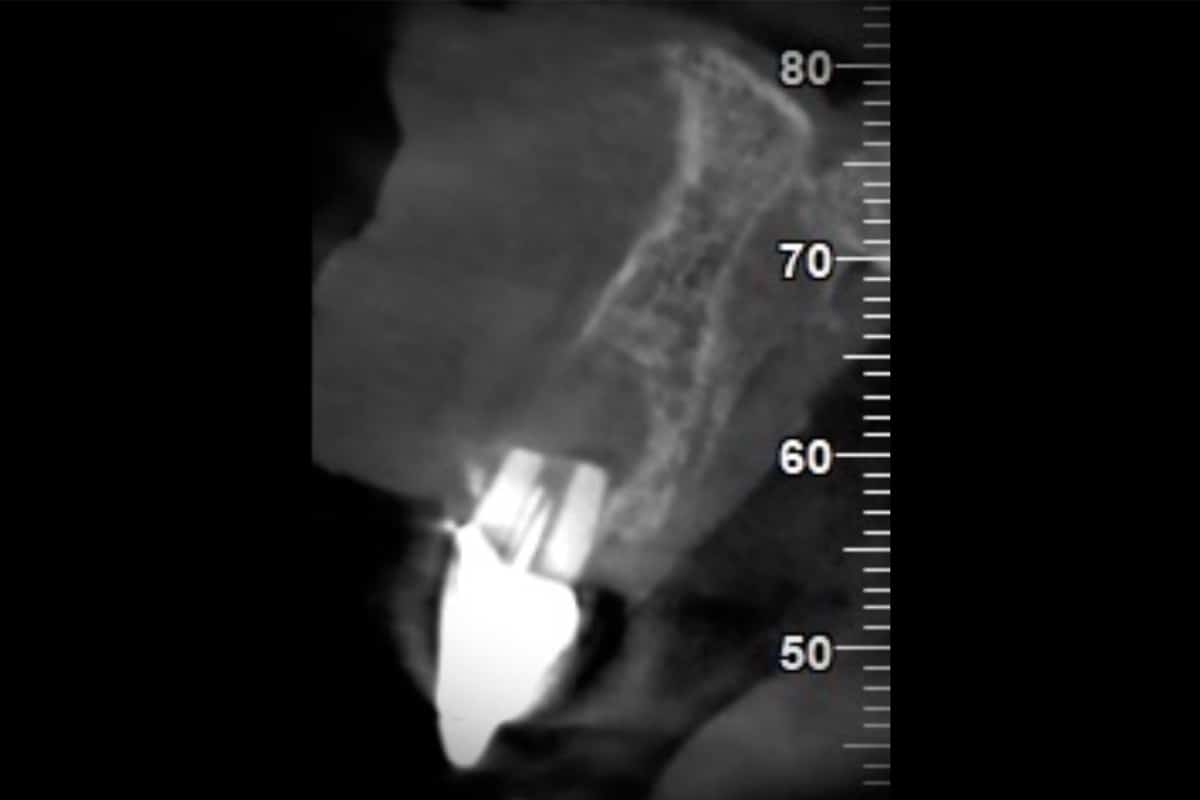
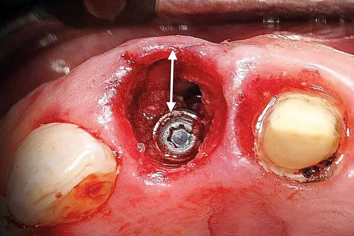
Clinical examination revealed tooth mobility and the presence of a fistula, while radiographic assessment demonstrated a buccal bone dehiscence with a radiolucent lesion, resulting in severe facial bone deficiency. Maintaining the ridge contour therefore represented a major challenge for immediate implant placement.
Treatment objectives:
- Immediate implant placement
- Reconstruction of the deficient buccal wall
- Preservation of the ridge contour
- Minimally invasive approach with open healing
Surgical Procedure
A treatment plan was established for guided immediate implant placement following tooth extraction.
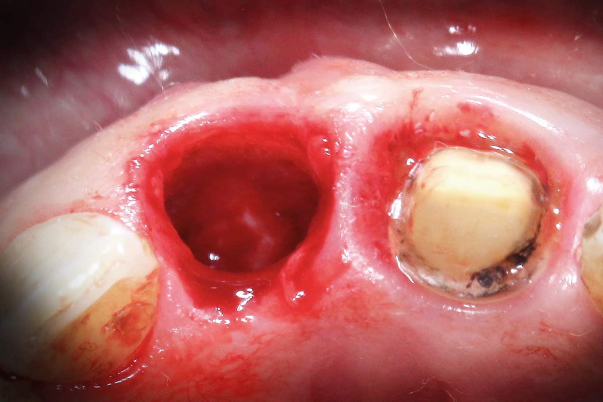
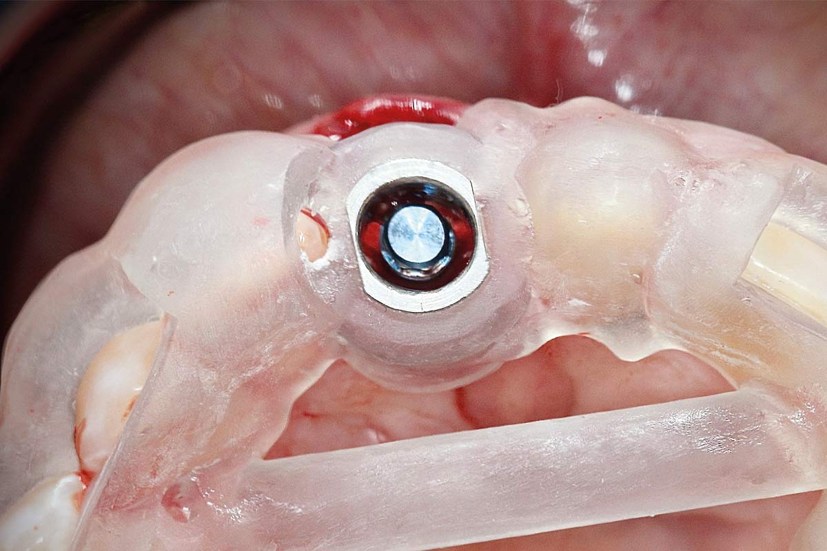
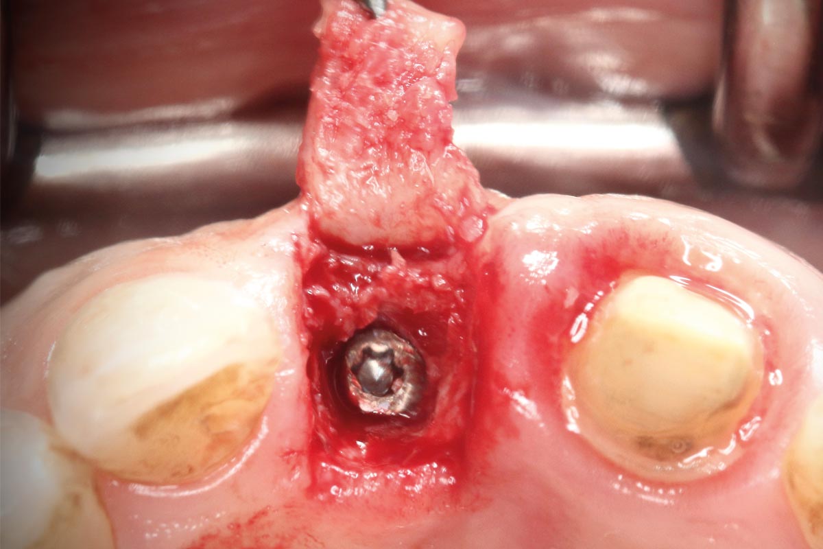
After atraumatic extraction and thorough socket debridement, the extent of the buccal dehiscence became fully apparent. A static surgical guide was used to achieve prosthetically driven implant positioning with adequate primary stability.
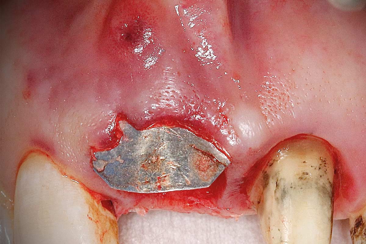
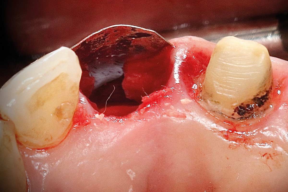
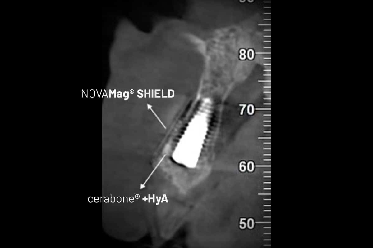
A subperiosteal tunnel was then prepared between the buccal dehiscence and the overlying soft tissue. NOVAMag® SHIELD was individually contoured and inserted into this space, reconstructing the missing buccal wall and creating a stable regenerative compartment.
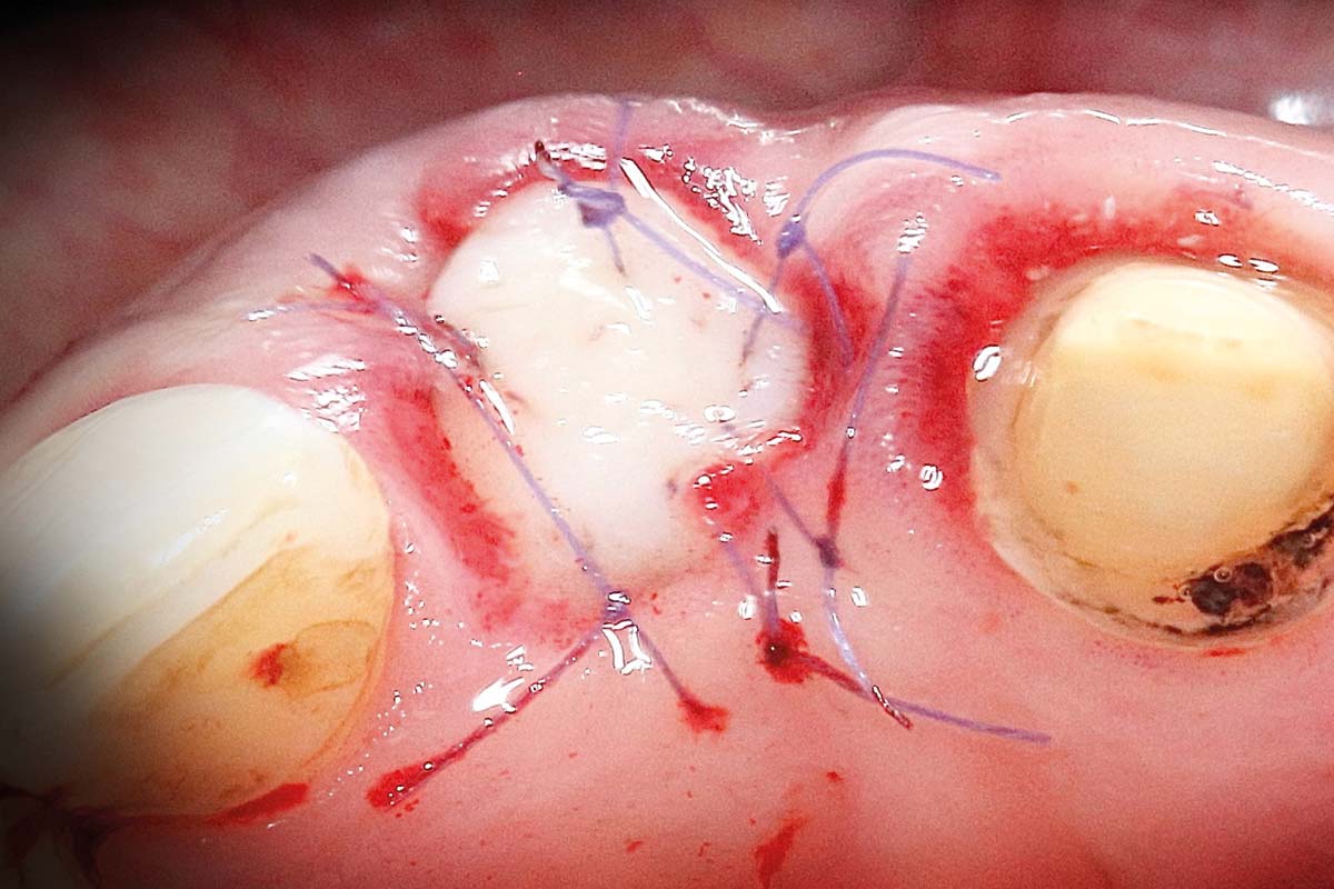
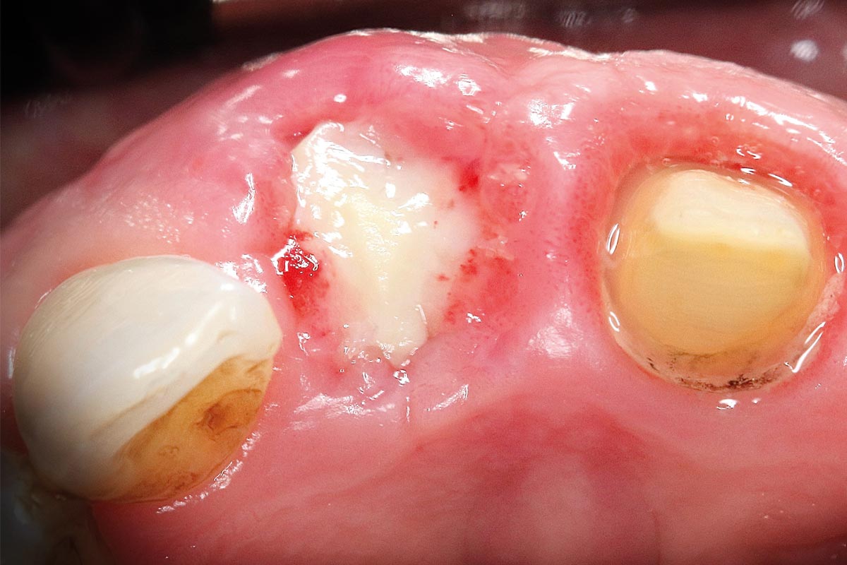
The space between the implant and the NOVAMag® SHIELD was augmented with cerabone® +HyA. The socket entrance was subsequently sealed with an autogenous palatal connective soft tissue graft, which was stabilized using sutures to support open healing.
Surgical workflow
- Atraumatic tooth extraction
- Guided immediate implant placement
- Preparation of a subperiosteal tunnel
- Placement of NOVAMag® SHIELD
- Augmentation with cerabone® +HyA
- Socket sealed with an autogenous palatal soft tissue graft and stabilized with sutures
Healing & Outcome
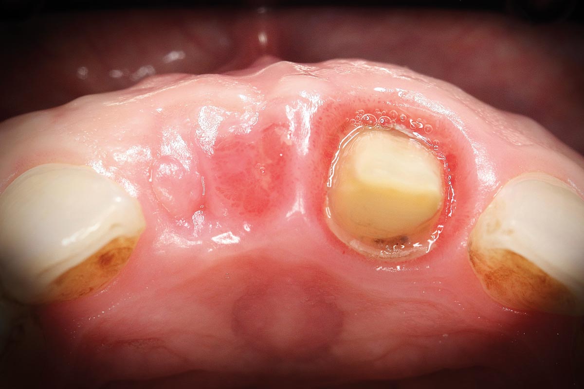
The soft tissue graft healed uneventfully, and the sutures were removed after 2 weeks. A 3-month healing period was allowed before re-entry.
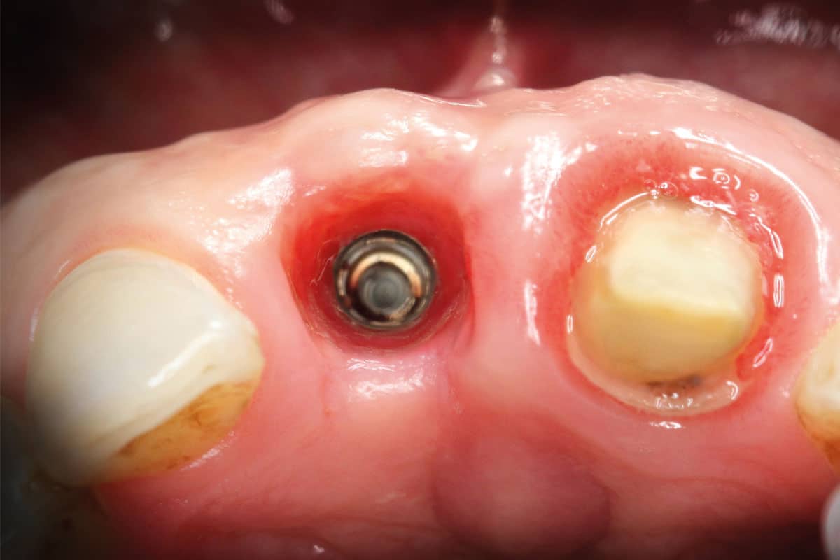
At re-entry, a healing abutment was placed. Clinical evaluation revealed excellent regeneration of the buccal bone, successful reconstruction of the facial contour, and healthy peri-implant soft tissue architecture with a favorable emergence profile, allowing progression to the prosthetic phase.
Postoperative radiographic evaluation demonstrated maintenance of the facial bone contour around the implant. As expected, NOVAMag® SHIELD was no longer radiographically visible due to its physiological degradation process and replacement by newly formed tissue.
Clinical outcome
- Uneventful healing
- Stable regenerated buccal bone
- Preserved ridge contour
- Healthy peri-implant soft tissue architecture
- Favorable emergence profile
- Implant successfully prepared for prosthetic restoration