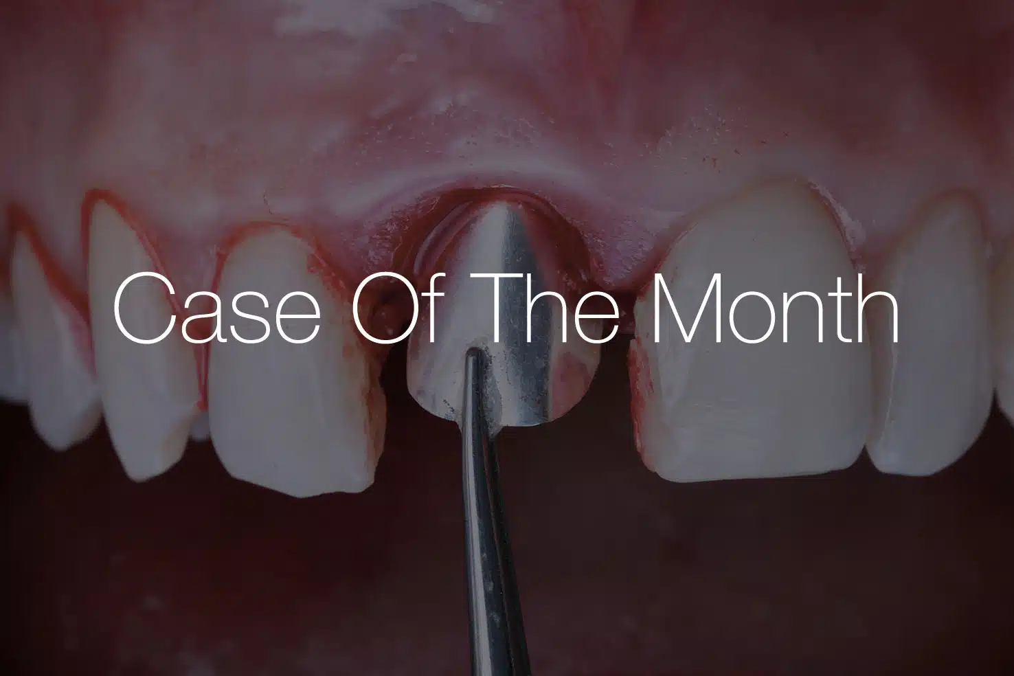
Socket management
Tooth extraction inevitably leads to remodelling of the alveolar ridge due to the loss of bundle bone. This physiological process may result in dimensional changes that can impact implant stability and the aesthetic outcome. Socket management and alveolar ridge preservation strategies aim to support the blood coagulum, maintain ridge volume, and preserve the integrity of the soft and hard tissues following tooth extraction.
TREATMENT APPROACH BASED ON
DEFECT MORPHOLOGY
The morphology of the buccal bone plays a key role in selecting the appropriate treatment approach. When the buccal wall is intact, immediate or early implant placement protocols may support natural healing of the blood coagulum and help maintain ridge dimensions. In cases with thin or compromised buccal bone, alveolar ridge preservation using bone substitute materials can help reduce resorption and preserve volume.
If the buccal plate is partially or completely missing, buccal plate reconstruction may be required as part of the socket management approach to recreate the contour of the ridge and enable optimal functional and aesthetic outcomes.

SOCKET TYPES IN
EXTRACTION SOCKET MANAGEMENT
Clinical decision-making in socket management is largely guided by the morphology of the extraction socket and the condition of the buccal bone.

Socket Typ 1
Buccal Bone Intact
Socket type 1 is characterized by an intact buccal bone plate and preserved socket walls following tooth extraction. The defect is fully contained, providing natural stability and favourable conditions for healing. In these cases, alveolar ridge preservation is typically straightforward, with stabilization of the blood coagulum allowing predictable bone regeneration and maintenance of ridge volume prior to implant placement.
Suggested approach:
Stabilization of the blood coagulum with collacone®
Socket Type 2
Compromised Buccal Bone
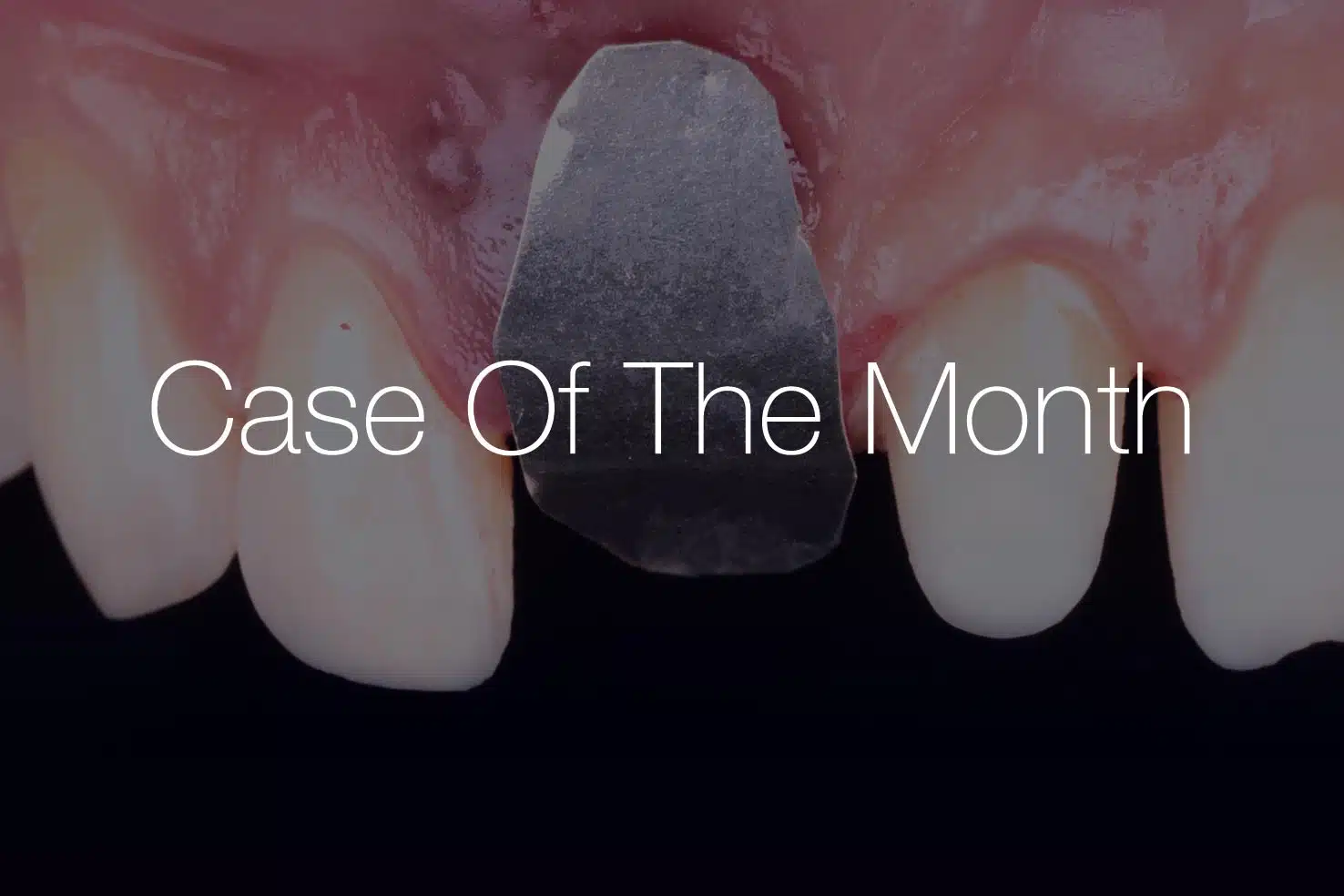
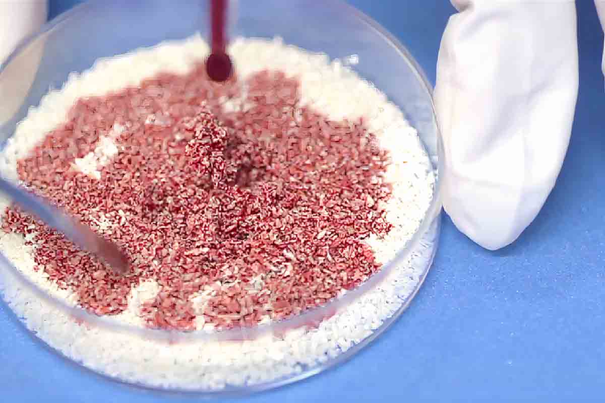
Socket type 2 presents with a compromised buccal bone, which may appear as dehiscence, fenestration, or an extremely thin buccal plate. In these situations, buccal wall preservation becomes critical. Grafting with a bone substitute is recommended to ensure adequate space maintenance, support the blood coagulum, and promote predictable bone regeneration as part of extraction socket management.
Suggested approach:
Buccal wall preservation with bone grafting using cerabone®+HyA & NOVAMag® SHIELD


Socket Type 3
Severe Buccal Bone Loss
Socket type 3 is characterized by severe loss of the buccal bone, often involving significant horizontal and/or vertical defects. In these situations, treatment focuses on ridge reconstruction, typically involving buccal plate reconstruction techniques to restore the ridge architecture and re-establish adequate bone volume for future implant placement.
Suggested approach:
Ridge reconstruction with early or delayed implant placement using cerabone®+HyA & NOVAMag® SHIELD
TREATMENT APPROACHES
SOCKET MANAGEMENT
Although physiological remodelling of bundle bone cannot be completely avoided, extraction socket management helps maintain the volume of the alveolus, facilitating future implant placement and improving long-term aesthetic outcomes. The application of bone substitute materials helps stabilize the blood coagulum, supports buccal wall preservation, and reduces ridge resorption.
Early implantation
After the natural healing of the extraction socket, an early implant placement procedure can be performed. According to the approach, the implant is placed after tooth extraction, before the bony regeneration of the socket has been completed. Typically, an early implantation is performed about 4–8 weeks after tooth extraction.
Delayed implantation
After complete healing and maturation of the extraction site, a delayed implant placement procedure can be carried out. In this approach, the implant is inserted only after full bony regeneration of the socket has occurred. Typically, delayed implantation is performed approximately 3–6 months after tooth extraction, depending on the extent of healing and the patient’s individual conditions.


Immediate implantation
Immediate implant placement refers to insertion of a dental implant directly after tooth extraction. Primary stability of the implant and the condition of the buccal bone are key factors. In favourable cases, immediate placement combined with appropriate socket management may support preservation of the alveolar ridge and aesthetic tissue contours.