
CLINICAL CASE
Dr. Kristian Grimm
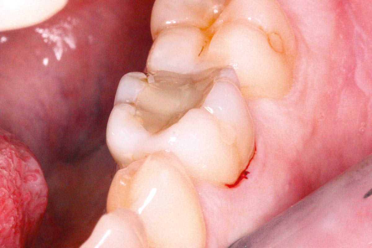
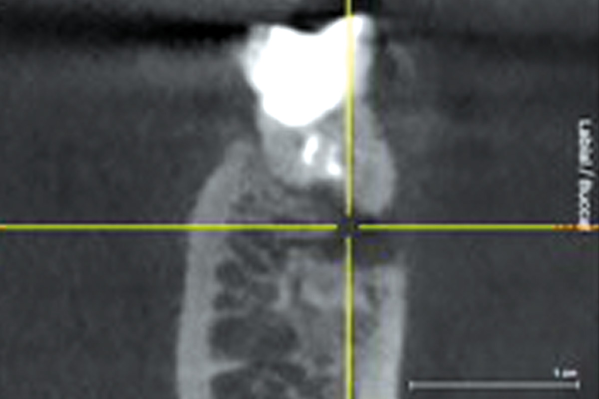
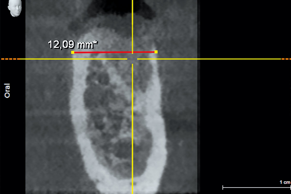
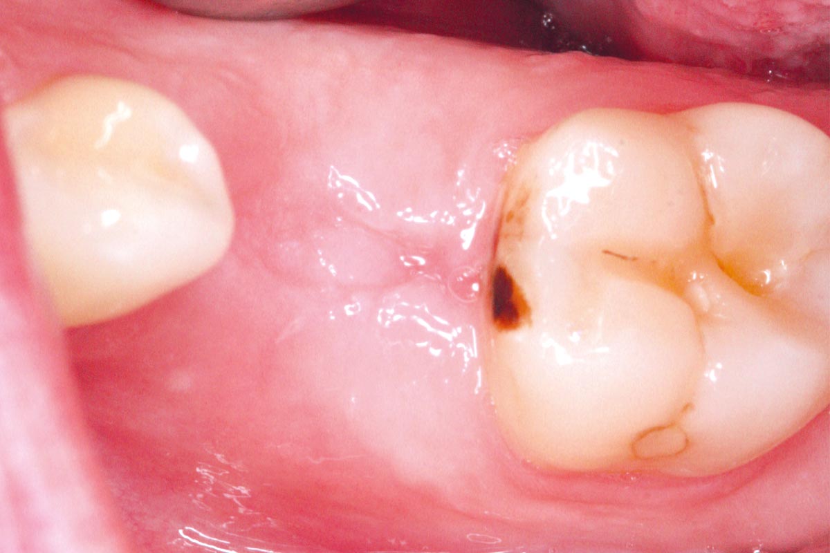
A 48-year-old female patient presented with persistent pain in the region of tooth 36. The symptoms occurred despite ongoing endodontic treatment. Clinically, there was pronounced tenderness to percussion, but no swelling or fistula formation. Cone beam computed tomography (CBCT) revealed an extensive periapical osteolysis at the distal root, along with a fine hairline crack in the same root.
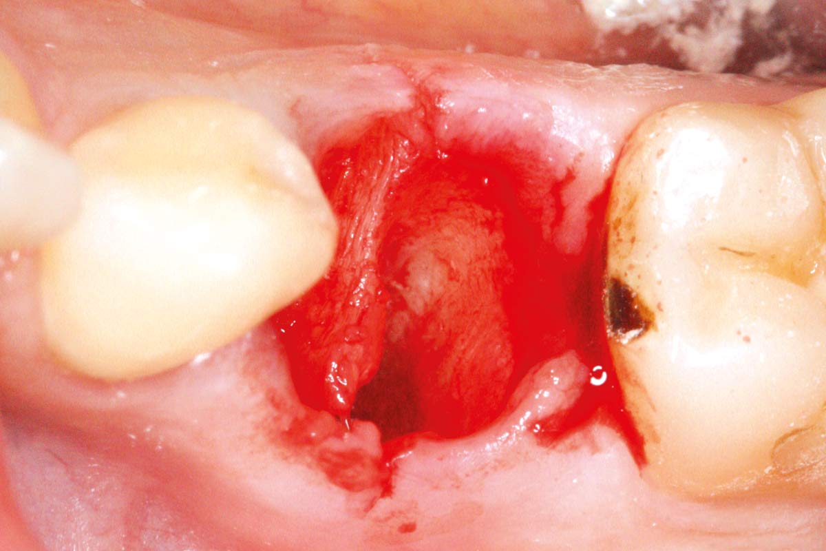
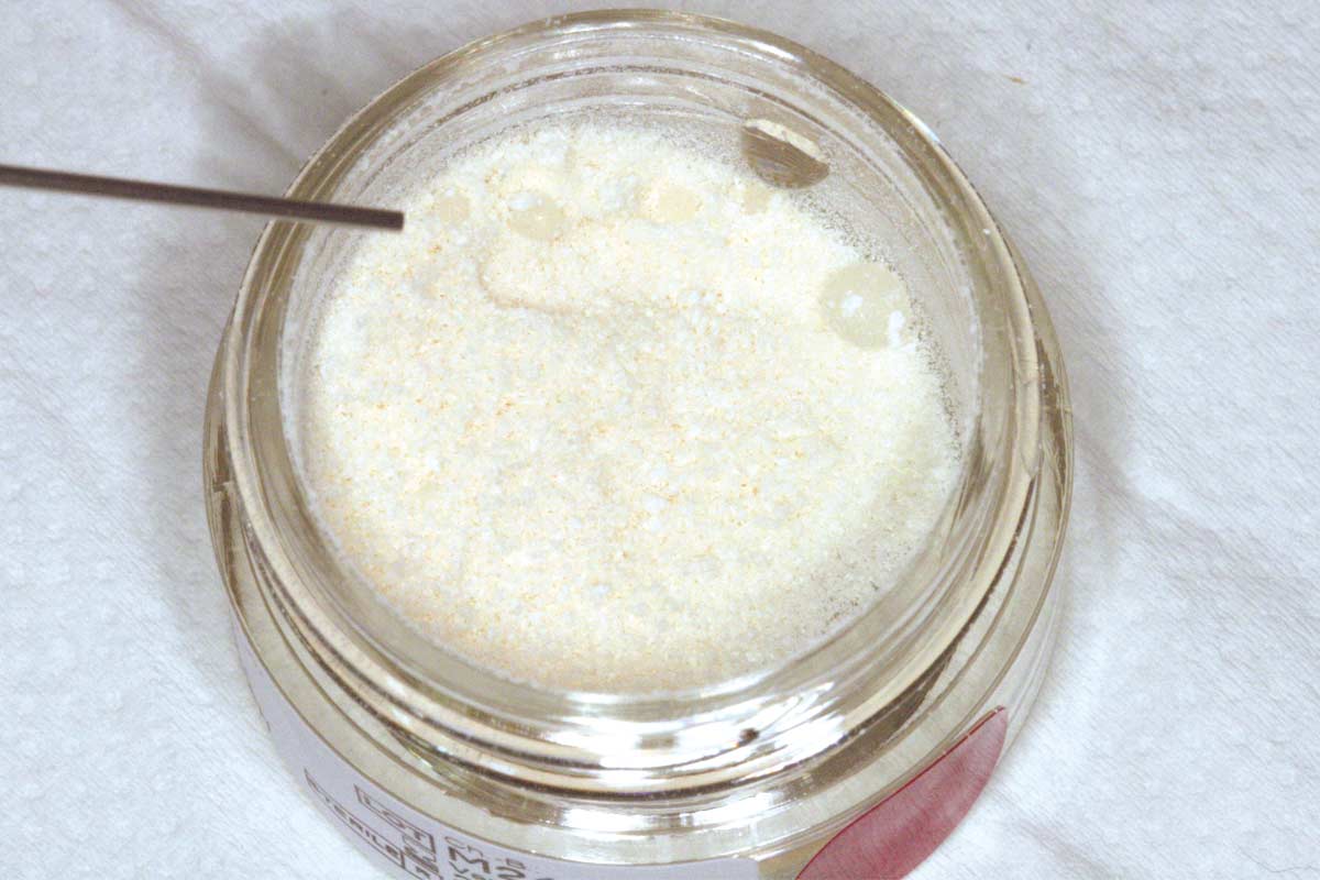
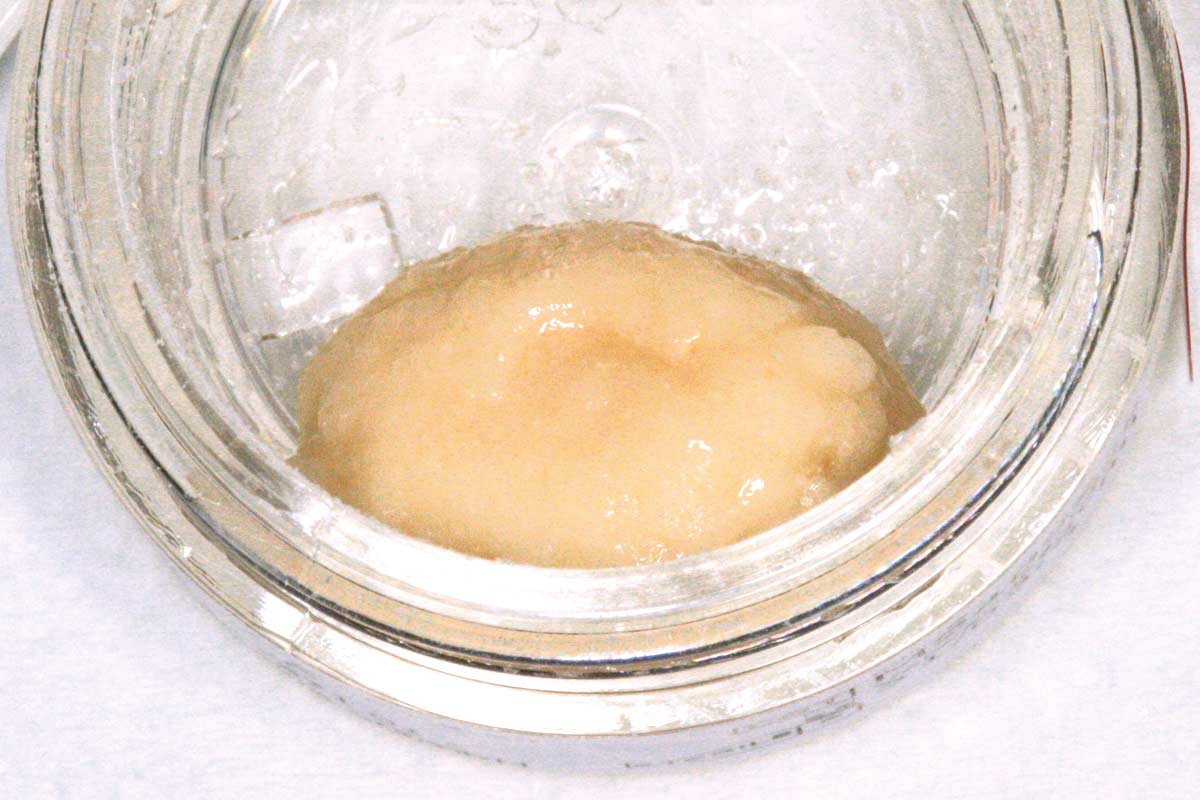
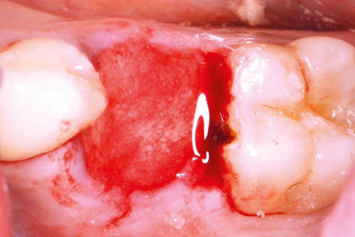
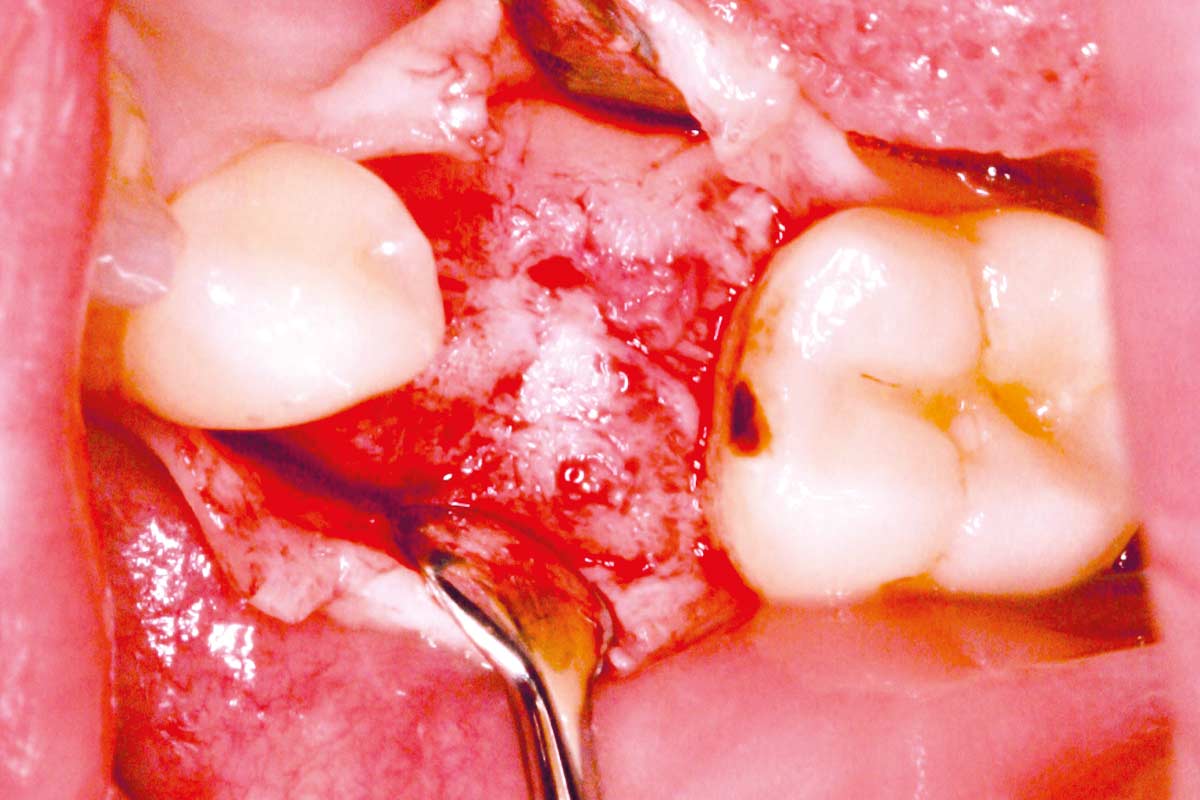
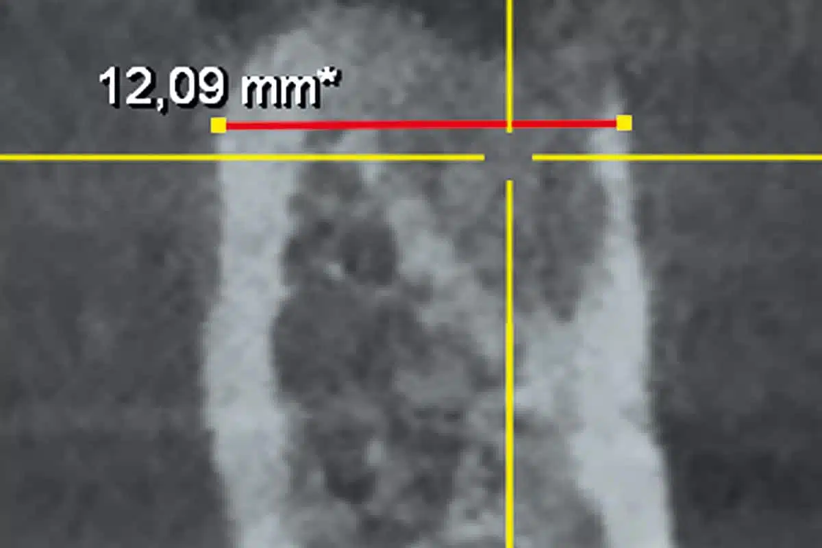
After thorough patient consultation regarding treatment options, the tooth was carefully extracted. Simultaneously, socket and ridge preservation were performed using cancellous maxgraft® +HyA – an allogeneic bone substitute material combined with hyaluronic acid. No membrane was used to cover the bone graft, only mattress suture. Postoperative healing was uneventful.
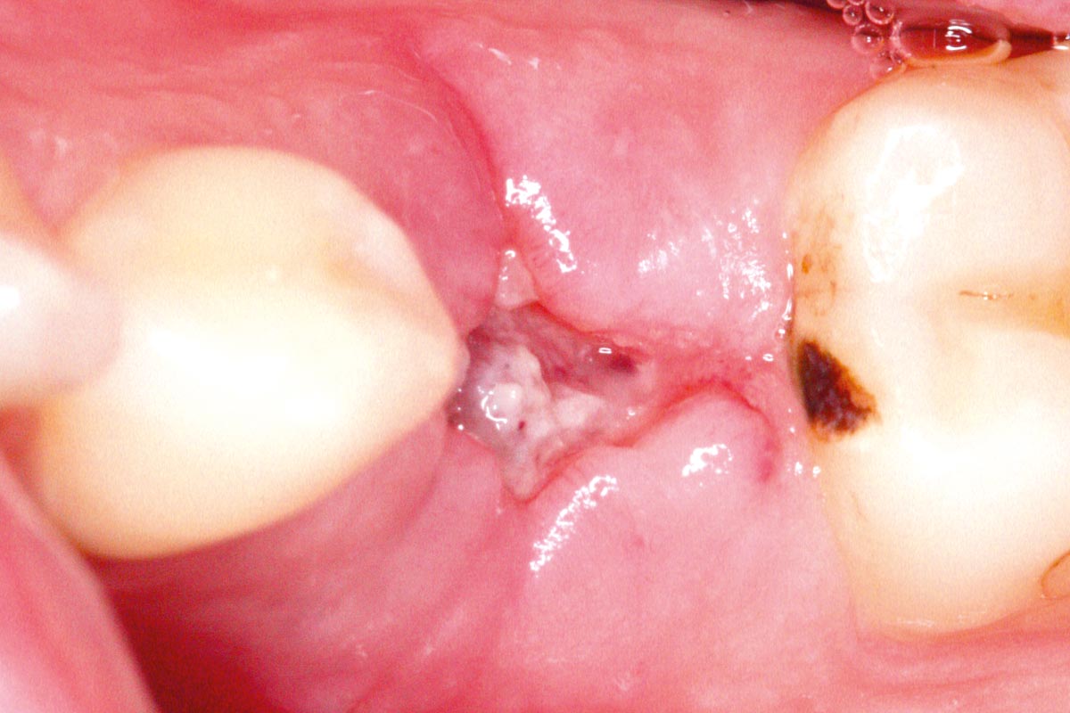
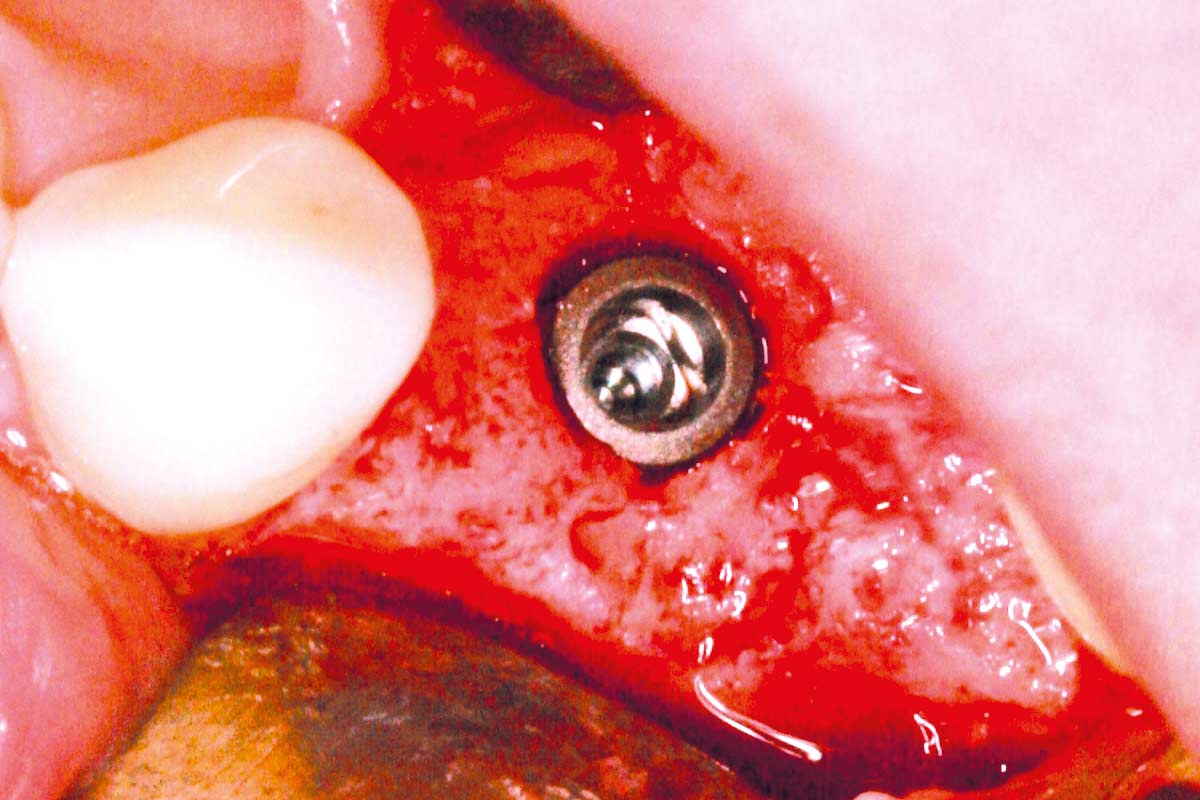
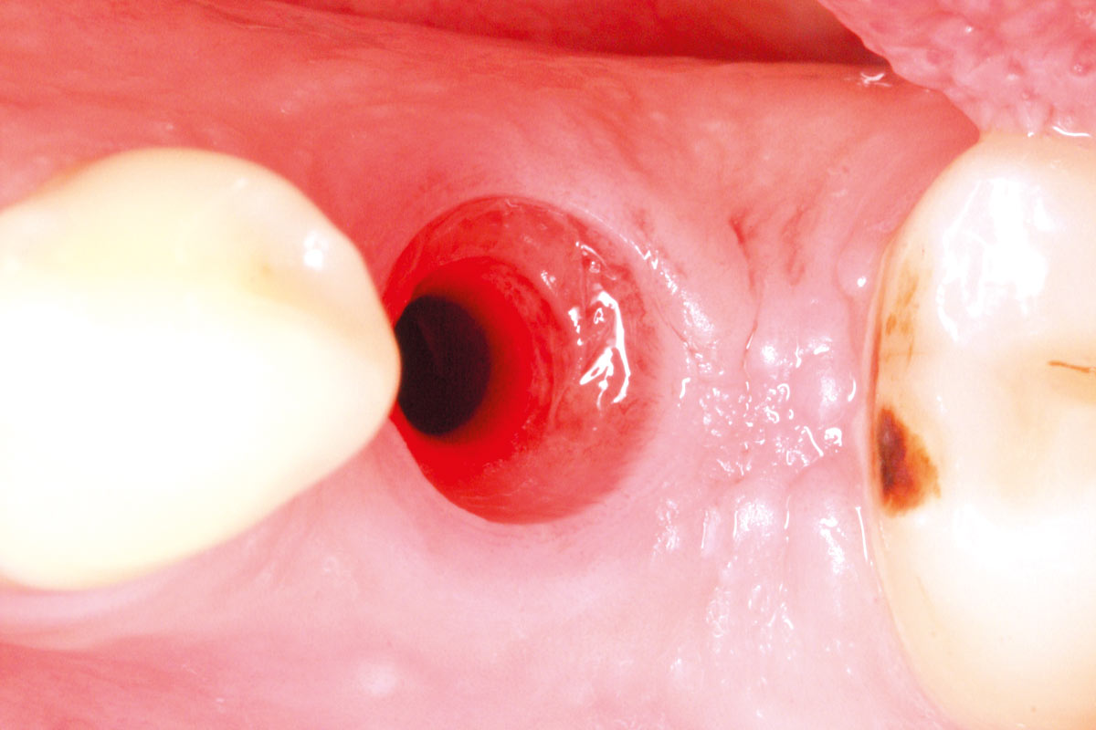
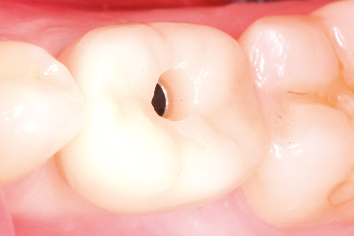
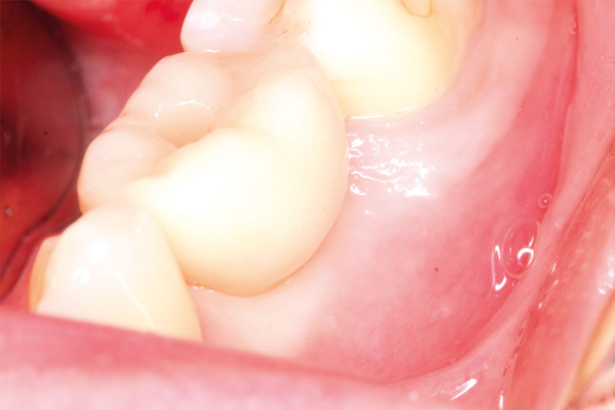
Ten weeks after extraction, an implant (diameter 5.2 mm, length 10 mm) was placed. The primary stability exceeded 35 Ncm, allowing for early prosthetic restoration. Eight weeks post-implantation, the final restoration was completed using a screw-retained crown, fabricated chairside with the CEREC system.
Conclusion:
This case highlights the importance of comprehensive diagnostics in patients with persistent symptoms during endodontic treatment. Early detection of a root fracture using advanced imaging enabled targeted treatment planning. The combination of socket preservation, timely implant placement, and digital chairside restoration resulted in a functionally and aesthetically successful outcome with minimal treatment time.