Study
Wen S-C, Barootchi S, Huang W-X, Wang H-L. J. Periodontol. 2019, Article in press
– Histology image with kind permission of Prof. Dr. med. W. Götz, Universitätsklinikum Bonn –
https://doi.org/10.1002/JPER.19-0142
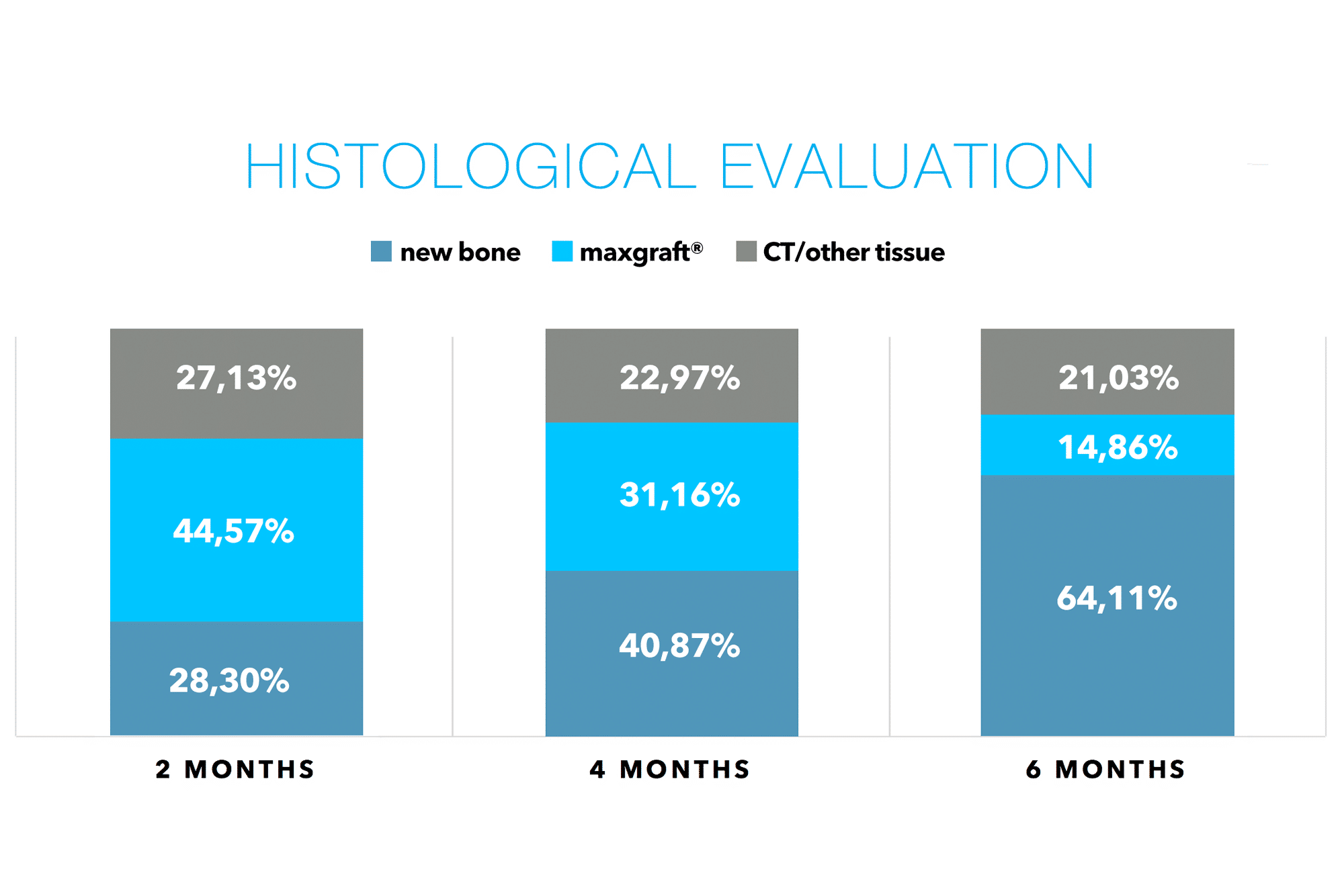
In the present study, the authors sequentially evaluated the healing of sockets augmented with a cancellous allograft (maxgraft®) and a d-PTFE membrane at 2, 4, and 6 months to compare the amount of vital bone, residual graft particles, and CT/other tissue.

Background: The objective of this study was to histologically evaluate and compare vital bone formation, residual graft particles, and fraction of connective tissue (CT)/other tissues between three different time points at 2‐month intervals after alveolar ridge preservation with a cancellous allograft and dense‒polytetrafluoroethylene (d‐PTFE) membrane.
Methods: Ridge preservation with a cancellous allograft and d‐PTFE membrane was performed at 49 extraction sockets (one per patient). Volunteers were assigned to implant placement at three different time points of 2, 4, and 6 months, at which time core biopsies were obtained. Histomorphometric analysis was performed to determine the percentages of vital bone, residual graft particles, and connective tissue/other non‐bone components, and subjected to statistical analyses.
Results: There was a statistically significant difference in the amount of vital bone at every time point from 28.31% to 40.87% to 64.11% (at 2‐, 4‐, and 6‐month groups, respectively) (P < 0.05). The percentage of residual graft particles ranged from 44.57% to 36.16% to 14.86%, showing statistical significance from 4 to 6 months (21.29%, P < 0.001), and 2 to 6 months (29.71%, P < 0.001), while there were no significant differences for the amount of CT/other tissue among the different time points.
Conclusions: This study provided the first histologic comparison of alveolar ridge preservation using a cancellous allograft and d‐PTFE membrane at three different time points. Extraction sockets that healed for 6 months produced the highest amount of vital bone in combination with the least percentage of residual graft particles, while similar results were observed for the fraction of CT/other tissues between the three time points.
Prof. Hom-Lay Wang


