CASE OF THE MONTH | 12/2024
Dr. Marko Blašković, HR
Blašković, M.; Butorac Prpić, I.; Aslan, S.; Gabrić, D.; Blašković, D.; Cvijanović Peloza, O.; Čandrlić, M.; Perić Kačarević, Ž. Magnesium Membrane Shield Technique for Alveolar Ridge Preservation: Step-by-Step Representative Case Report of Buccal Bone Wall Dehiscence with Clinical and Histological Evaluations. Biomedicines 2024, 12, 2537. https://doi.org/10.3390/biomedicines12112537
Original title: Alveolar ridge preservation with NOVAMag® membrane: Buccal Bone Wall Dehiscence with the Shield technique
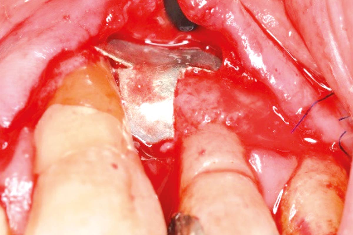
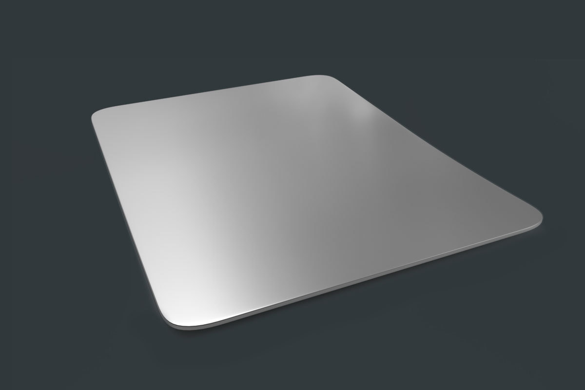
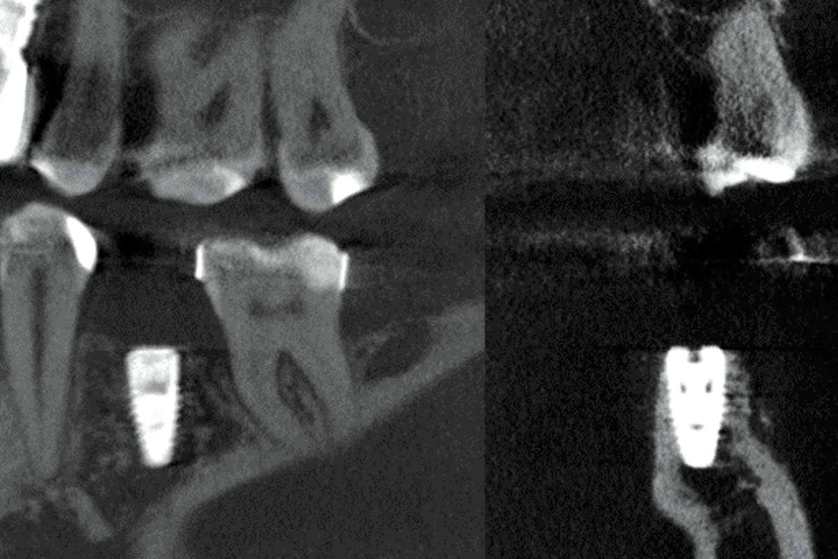
A 44-year-old female patient in good general health with no chronic conditions presented with gingival recession around tooth 24, which had a vertical fracture line and a periapical lesion, necessitating extraction due to severe buccal bone wall loss. Cone beam computed tomography confirmed previous endodontic treatment, a periapical lesion, and complete destruction of the buccal bone wall. After tooth extraction, an intrasulcular incision was made to elevate a flap, facilitating access to the defect area. The NOVAMag® SHIELD was positioned beneath the periosteum, covering the entire defect area and extending an additional 3 mm onto the adjacent intact bone in the apical, mesial, and distal directions to ensure optimal stabilization and coverage. A bone graft mixture of autogenous bone and cerabone® filled the socket, with the membrane secured over the crest, supporting open healing. Then, the NOVAMag® SHIELD was bent and rolled over the crestal part of the ridge and tucked below the palatal flap. This approach maintains the membrane’s placement while allowing for open healing, as the membrane is left exposed for healing by secondary intention. Follow-up visits at 2, 3, and 6 months showed uneventful healing, with no complications. After six months, an implant was placed, followed by a free gingival graft to improve soft tissue thickness. At implant placement, a biopsy was harvested for scientific purposes. Four months post-OP, the patient began the final prosthetic restoration process, achieving stable soft tissue contours and satisfactory esthetic outcomes. Histological analysis confirmed healthy, mature bone formation with visible osteocytes and active osteoblasts, indicating successful integration and bone regeneration.
The „Case Of The Month“ highlights every month a clinical case, which distinguished itself by the clinical results or the treatment concept in combination with the applied botiss biomaterials. The selection of the case is based on content relevance and quality of the documentation.


