CLINICAL CASE
Dr. Marko Blašković
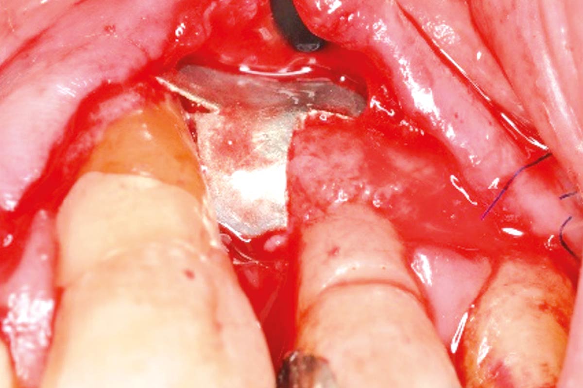
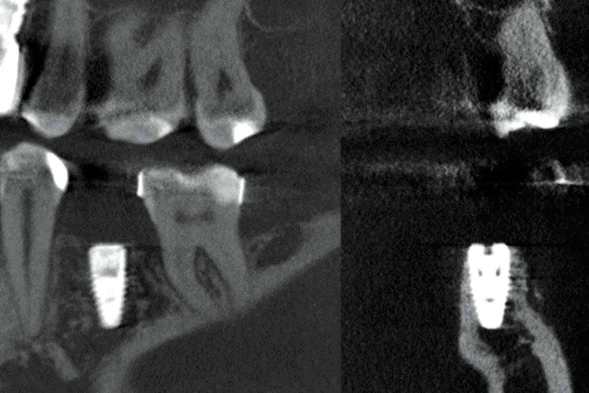
Cone beam computed tomography (CBCT) of tooth 12 (FDI notation system) revealed bone deficiency in both horizontal and vertical directions. A complete mucoperiosteal flap was raised and the site intended for augmentation was exposed. cerabone® and a small amount of locally harvested autogenous bone were mixed together and used to augment the defect. NOVAMag® membrane was used to separate the defect site from the overlying soft tissue. The membrane was secured to prevent slippage using resorbable NOVAMag® fixation screws. To achieve an optimal soft tissue profile, mucoderm® was also placed over the membrane. The clinical outcome after 3 months was satisfactory, with the labial and oral mucosas being close together, and soft tissue seen to be regenerated. After a total of 5 months of healing, CBCTs show bony healing in vertical and vestibulo-ral dimensions.