CASE REPORT
Original title: First Clinical Case Report of a Xenograft–Allograft Combination for Alveolar Ridge Augmentation Using a Bovine Bone Substitute Material with Hyaluronate (Cerabone® Plus) Combined with Allogeneic Bone Granules (Maxgraft®).
Kloss, F.R.; Kämmerer, P.W.; Kloss-Brandstätter, A. J. Clin. Med. 2023, 12, 6214.
https://doi.org/10.3390/jcm12196214
This case report demonstrates Guided Bone Regeneration for later implant placement in the aesthetic zone in a 34-year-old patient using a combination of freeze-dried bone allograft and pure bovine bone mineral with hyaluronate.
INITITAL SITUATION
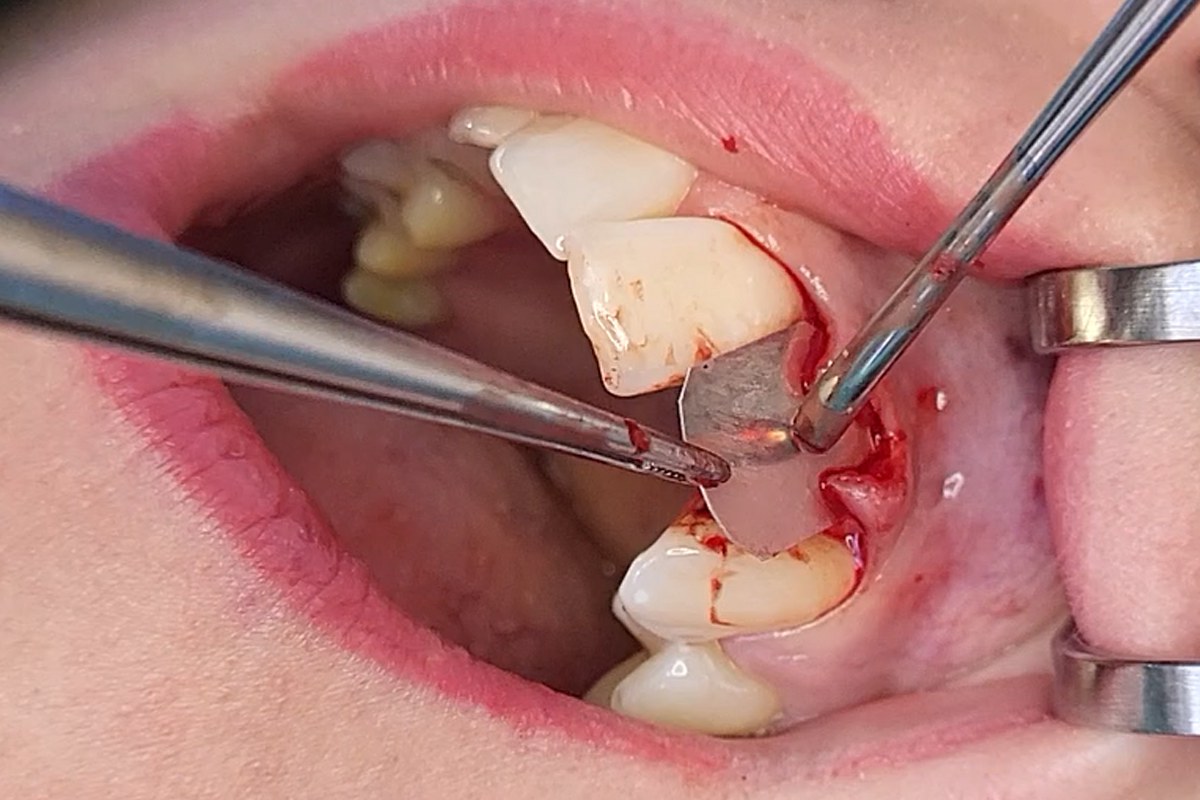
Tooth 21 suffered from a trauma 15 years ago and was mobile. Panoramic x-ray demonstrated a periradicular lesion, and the clinical examination revealed the presence of a buccal fistula. Therefore, the tooth was extracted. A slight lack of buccal bone was evident; however, the soft tissue situation was inflammation-free, and a broad zone of keratinized gingiva was visible. Otherwise, the patient was healthy without any symptoms of periodontitis.
TREATMENT APPROACH
CBCT analysis showed a transverse bone defect of the alveolar ridge with a residual ridge width of 2–3 mm. Based on the geometry of the defect a two-stage Guided Bone Regeneration (GBR) procedure was planned using bovine bone mineral with hyaluronate (cerabone® plus) mixed with allograft bone substitute from human donor bone (maxgraft® cancellous granules) in conjunction with a porcine pericardium membrane (Jason® membrane). Implant placement was planned six months post-augmentation.
OUTCOME
The healing was uneventful without signs of infection, wound dehiscence, graft exposure or other postoperative complications. Six months post-surgery, re-entry and implant placement were performed. The bleeding bone indicated satisfactory osseous transformation and good blood supply to the graft. Moreover, the biomaterials mix of xeno- and allograft was well integrated into the recipient site, and the formerly lacking bone volume was sufficiently restored. A titanium implant (Medentika; Straumann Group) was placed into the regenerated area, which was uncovered three months later to connect a screw-retained crown.
FOLLOW-UP
Three years postoperatively the soft tissue situation was stable without signs of mucositis at the implant site. The panoramic radiograph also revealed excellent osseointegration of the implant. No signs of bone loss at the implant were detectable.
CONCLUSION
The biomaterials combination of bovine bone mineral with hyaluronate and bone allograft provided optimal conditions for successful bone regeneration. cerabone® plus offers volumetric stability, while the allogeneic bone promotes fast natural bone regeneration. Moreover, thanks to its sticky properties after hydration, cerabone® plus makes defect augmentation effective and permits precise application of the granules, as well as quick defect contouring.


